
Research Article: Journal of Drug and Alcohol Research (2026) Volume 15, Issue 8
New Hypotheseses on Pathogenesis of Depression in Parkinson’s Disease
Saumya Mathew1, Sibi Joseph1, Lourdes de Fatima Ibanez Valdes2 and Humberto Foyaca Sibat2*2Department of Internal Medicine, Nelson Mandela Academic Hospital, Walter Sisulu University, South Africa
Humberto Foyaca Sibat, Department of Internal Medicine, Nelson Mandela Academic Hospital, Walter Sisulu University, South Africa, Email: humbertofoyacasibat@gmail.com
Received: 13-May-2026, Manuscript No. JDAR-26-192699; Editor assigned: 15-May-2026, Pre QC No. JDAR-26-192699 (PQ); Reviewed: 29-May-2026, QC No. JDAR-26-192699; Revised: 17-Aug-2026, Manuscript No. JDAR-26-192699 (R); Published: 24-Aug-2026, DOI: 10.4303/JDAR/236524
Abstract
Objectives: This study’s primary goal is to review the most recent data that has been published in the medical literature about the pathophysiology of depression in Parkinson’s disease.
Methods: To discover papers pertaining to new information on the pathophysiology of depression in Parkinson’s disease, a thorough search of the medical literature was conducted using the databases PubMed/ MEDLINE, Scopus, and Embase. We used PRISMA standards to search the medical literature between January 1, 2000, and January 31, 2026. The following terms were used to search Scopus, Embase, and PubMed Central: “Pathogenesis of depression,” “Pathogenesis of Parkinson’s disease,” “Microbiota,” “Gutbrain axis,” “Dysbiosis,” “Limbic system,” “Major depressive disorders,” “Dopaminergic dysfunction,” “Network-level disturbances,” and “Vagal Nerve (VN)”.
Results: 1563 articles were first found using a literature search. 652 publications remained after 911 duplicates were eliminated based on title and abstract review. Of these, 174 papers examined the pathophysiology of depression in Parkinson’s Disease (PD), as 478 did not fit the inclusion/ exclusion criteria. Twelve of these publications offered new insights into the pathophysiology of Parkinson’s disease. Nevertheless, a quality evaluation showed that none of the included papers offered novel theories.
Conclusions: Recent research has shown that depression in Parkinson’s disease is associated with dopaminergic dysfunction, network-level disruptions, dysbiosis, and neurodegeneration. processes. To the best of our knowledge, this review is the first to concentrate on new facets of the pathophysiology of depression in Parkinson’s disease. Additionally, it presents fresh theories regarding how the limbic system and other brain areas are impacted by the microbiome
Keywords
Introduction
Parkinson’s Disease (PD) is a neurological movement illness that progresses over time. Alpha-synuclein (a-Syn)- rich aggregates build up and dopaminergic neurons in the posterolateral part of the Substantia Nigra (SN) pars compacta are lost. It manifests as non-motor symptoms such sadness, cognitive impairment, anxiety, and olfactory dysfunction in addition to motor symptoms like bradykinesia, stiffness, and tremor [1].
Additionally, it is acknowledged as an age-related neurodegenerative illness for which there is presently no treatment [2,3].
It affects about 2% of the global population aged 65 years and older. The increasing prevalence and earlier onset contribute to a significant socioeconomic burden [4]. This underscores the critical need for well-designed animal studies to recapitulate both PD’s molecular hallmarks and behavioural symptoms, and to elucidate its pathogenesis and novel therapeutic approaches.
The progressive loss of dopamine-producing neurons in the substantia nigra, a part of the brain crucial for motivation and movement, is the fundamental pathology of Parkinson’s disease. Dopamine levels in the basal ganglia decrease as these neurons deteriorate, resulting in both motor and nonmotor symptoms. The buildup is a crucial pathogenic aspect of this process. of aberrant protein clumps called Lewy bodies, which are linked to persistent neuronal dysfunction and cell death and contain misfolded alpha-synuclein [5].
Bradykinesia, stiffness, postural instability, and resting tremor are typical motor signs of Parkinson’s disease. But non-motor symptoms including melancholy, constipation, sleep issues, and cognitive decline are becoming more widely acknowledged as essential elements of these symptoms frequently appear years before motor signs and may occur throughout the prodromal phase. As a result, non-motor symptoms are crucial in identifying those who may develop Parkinson’s disease [6,7].
Motor manifestations by several years, and therefore play a critical role in the early identification of individuals at risk of developing PD [6,7].
Furthermore, functional deterioration and a lower quality of life are significantly influenced by non-motor symptoms. Particularly significant are psychiatric symptoms, especially apathy, anxiety, and depression [7].
Depression, as defined by the Diagnostic and Statistical Manual of Mental Disorders (DSM-5-TR), is characterised by symptoms including depressed mood, anhedonia, feelings of guilt or worthlessness, impaired concentration, appetite changes, sleep disturbances, and recurrent thoughts of death [8].
In Parkinson’s disease, however, the clinical presentation of depression often differs from that seen in primary Major Depressive Disorder (MDD). Standard diagnostic criteria remain applicable.
However, depressive symptoms in PD less commonly involve overt sadness, guilt, and suicidal ideation. Instead, they more often present as cognitive impairment, reduced motivation, and difficulties with concentration [9].
Suicidal ideation, and more commonly present with cognitive impairment, reduced motivation, and difficulties with concentration [9].
This distinction suggests that depression in Parkinson’s disease may not merely represent a psychological response to chronic illness, but rather a manifestation of the underlying neurodegenerative process itself. Disruption of dopaminergic, serotonergic, and noradrenergic pathways, alongside the dysfunction of limbic and fronto-striatal circuits involved in mood regulation, it provides a biological basis for this altered clinical presentation [5].
Weintraub and associates from the Psychiatry and Neurology Departments at the University of Pennsylvania examined Neuropsychiatric (NP) signs of Parkinson’s disease in 2022. They concurred that these symptoms had the potential to cause just as much handicap as motor ones. To enhance the management of NP manifestations in Parkinson’s disease, they suggested specialized training, more research, increased awareness, and the creation of creative care models.
Perceptual and cognitive problems, such as psychosis; motivational symptoms, such as apathy and impulse control difficulties; and, most frequently, emotional disturbances, such as anxiety and sadness, are the hallmarks of this complex NP illness [10].
Materials and Methods
A comprehensive search of the medical literature in the PubMed/MEDLINE, Scopus, and Embase databases was conducted to identify articles reporting novel information on GA and diagnostic procedures.
From 01st, January 1989 to 30th, November 2025, we searched the medical literature, following the PRISMA guidelines. The authors searched the scientific databases, Scopus, Embase, Medline, and PubMed Central using the following searches: “Gait apraxia” OR “Walking skills” OR “Apraxia of postural transitions” OR “Diagnostic tools” OR “Limb apraxia”, OR “Pathophysiology of apraxia” OR “Treatment/Management of apraxia” OR “Corticobasal syndrome” OR “Normal pressure hydrocephalus”.
The systematic review performed in this study followed the guidelines recommended by PRISMA (2020 statement).
Search strategy
We searched PubMed to access MEDLINE for articles published in English between Jan 1, 2015, to June 13, 2021, using the medical subject headings search terms: “Parkinson and depression”; “Parkinson and (psychosis or hallucination or delusion)”; “Parkinson and anxiety”; “Parkinson and (impulse control disorder or dopamine dysregulation syndrome)”; and “Parkinson and apathy”. The systematic review filter was used to narrow the search. A systematic search was conducted across the following databases: PubMed Central/MEDLINE, Scopus, and Embase to identify publications on the pathophysiology of GA, management, and diagnostic procedures.
Only articles published in English, Spanish and Portuguese were selected. Editorials, preclinical studies, and conference proceedings were excluded.
Selection of study
All investigators (SM, SJ, LdeFIV, and HFS) separately reviewed the abstracts and titles of the selected publications and independently reviewed the full-text versions of the identified articles to determine their eligibility for inclusion. Furthermore, publications without a clear protocol for confirmatory diagnosis, papers lacking analysis, publications with incomplete data, or those that did not mention the exact number of patients or pathogenesis of depression and PD were not considered for inclusion.
Selection criteria
The following criteria were included: Articles with detailed pathogenesis and/or drug management. Clinical features of LIS and demographic information.
Exclusion criteria were applied:
• Inaccessibility to full text
• Articles with unclear pathogenesis
• Lack of relevant clinicopathological data
• Non-original studies (i.e., editorials, letters, conference proceedings, book chapters)
• Animal model studies
• non-/Spanish/Portuguese/English studies.
The papers that were not thoroughly assessed were removed.
Data extraction and quality assessment
The studies’ quality assessment, including applicability concerns and risk of bias, was categorised as good, poor, fair, or reasonable, in agreement with the National Institutes of Health Criteria and the Quality Assessment of Diagnostic Accuracy Studies version 2 (QUADAS-2) evaluation. Quality evaluation was made separately by both authors, and disagreements were resolved by scientific discussion and final agreement.
Data collection, extraction, and bias assessment
Before being cited to gather pertinent data for the review, two authors revised all abstracts and titles that met the inclusion criteria. Data on the author’s name, age, year of publication, country of information source, kind of study, total number of cases included, and pathophysiology of depression in PD patients were gathered for each publication chosen for the review. An Excel spreadsheet was updated with the chosen data from articles that qualified.
Outcome measures
Our plan was to select the most relevant publications on the pathogenesis of depression in PD. This investigation also identified disorder associated with the enteric nervous system such clinical repercussion of dysbiosis.
Statistical analysis
Statistical analysis was performed using XLSTAT (add-on for Microsoft Excel, version 2021.4.1, Addinsoft SARL and RStudio.
Results and Discussion
Literature search
Searching the literature retrieved 1563 articles. After reviewing the titles and abstracts, 911 duplicate publications were removed, leaving 652 publications selected. After applying the inclusion/exclusion criteria, 478 articles were excluded; therefore, 174 studies investigated the pathogenesis of depression in PD. Looking for novel information on the pathogenesis of PD, 12 manuscripts were selected. For quality assessment, no articles that delivered new hypotheses was included (Figure 1).
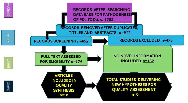
Figure 1: PRISMA flow diagram with included publications
To provide an accurate assessment of this search, the authors used a QUADAS-2 evaluation to determine that the risk of bias was low/moderate for almost all publications. The diagnosis of GA was made in all patients according to the current proposed criteria. The authors considered the substantial technical differences observed across diagnostic protocols used in several studies.
Noticeably, in some publications, small and mixed cohorts were processed, including different types of diagnostic procedures, leading to fewer GA cases examined under the same protocol.
Comments and final remarks
Based on well-designed longitudinal and cross-sectional studies, some authors have concluded that the severity and prevalence of NP symptoms and signs increase over time [11].
And those manifestations can present in isolation but are often multimorbid and result from complex mechanisms [10].
Last year, Ellul and collaborators showed that biomarkers of other neurocognitive disorders, such as amyloid-beta and phosphorylated-tau, can predict cognitive and affective trajectories in PD. Therefore, using a multi-modal panel of prognostic markers might accurately predict cognitive and affective outcomes in PD [12].
Preliminary investigations have revealed that decreased levels of amyloid beta [13] and alpha-synuclein [14] decreased levels of amyloid beta [13] and alpha-synuclein [14] in the Cerebrospinal Fluid (CSF) are directly associated with attention and memory impairments, as well as increased depression in people with PD [15,16].
Equally, affective disturbance in early PD has been shown to predict intense affective disorder over disease duration,including PD-associated depression [17] and overall quality of life [18].
Both increased CSF phosphorylated-tau181 and reduced amyloid-beta levels significantly predicted membership in the cluster with more pronounced cognitive/affective dysfunction (cluster 2) at 5-year follow-up. Both are biomarkers of degenerative disorders like Alzheimer’s Disease (AD) and Parkinson’s Disease (PD). AD is associated with hyperaccumulation of both amyloid beta and phosphorylated tau [19].
High CSF concentrations of phosphorylated-tau and amyloid-beta aggregates are consistently reported in de novo PD [20,21]. The same pathology can be observed in PD.
Neurodegenerative Disorders (NDDs) as AD, PD, and ALS have complicated, multiple pathophysiologies. According to recent research, the Microbiota-Gut-Brain Axis (MGBA) is crucial for the onset and progression of NDD, particularly in PD patients. Through neurological, endocrine, immunological, and metabolic processes, the gut microbiota and the brain are connected by the MGBA, a two-way network. Recognizing these differences is essential, as depression in PD is frequently underdiagnosed and undertreated, despite its significant impact on morbidity, disease progression, and quality of life. Depression in PD is often under-recognised and undertreated, despite its substantial impact on morbidity, disease progression, and quality of life.
A clearer understanding of the underlying pathophysiological mechanisms may help identify depression earlier and enable more targeted therapies. In the end, this could lead to better patient outcomes by informing earlier detection and more focused therapeutic approaches. This thorough review reveals that only five papers that examine the pathophysiology of depression in Parkinson’s Disease (PD) have been published in 2026. Several recurrent themes- particularly those that were connected-emerged despite the small number of studies. to dopaminergic dysfunction, alterations in brain network connections, and the connection between depression and other non-motor symptoms such sleep difficulties and cognitive impairment.
Parkinson’s Disease (PD) is the clinical manifestation of loss of dopaminergic neurons in the posterolateral aspect of the pars compacta of the Substantia Nigra (SN) and the accumulation of alpha-Synuclein (a-Syn)-rich aggregates, presenting both motor and non-motor symptoms. Recently, Kondrataviciute and collaborators documented that the virus-mediated a-Syn overexpression in a rat model recapitulates progressive dopaminergic neurodegeneration in the SN, leading to motor deficits, while the non-motor phenotypic profile remains poorly characterised. They investigated the behavioural consequences of targeted Adeno-Associated Virus (AAV)-mediated hyperactivation of aggregate-prone mutant A53T a-Syn within the rat SN bilaterally [22]. These authors confirmed that within six weeks, a dopaminergic neurodegeneration happened, along with progressive motor impairments characterised by reduced locomotion as early as 3 weeks post-AAV injection and concluded that animals overexpressing A53T a-Syn exhibited reduced responsiveness to palatable stimulation and bilateral A53T a-Syn overexpression induces depression-like behaviour, providing a remarkably important tool for investigating the pathogenesis and therapy of non-motor affective symptoms in PD which has been supported by other investigators [23].
Common neuropsychiatric presentations in PD The most common non-motor features in PD are the Neuropsychiatric (NP) symptoms and signs. Furthermore, an increased incidence, prevalence rates and symptoms preceding the presentations of motor signs have been reported [24,25].
At the early phase of PD, some NP signs and symptoms may happen in isolation, although they quite often occur, like psychosis and depression, depression with anxiety, apathy overlaps with depression, cognitive impairment and can lead to complex impulse control disorders [26].
At the late stage, individual neuropsychiatric symptoms and signs can be predicted by comorbid NP symptoms and signs [25].
Despite investigations done, it is still a controversial issue whether NP manifestations of PD should be accepted as unique to the disorder or as pseudo-specific. On the other hand, it’s unclear whether the affective manifestations identified in PD patients are different from those reported in the general population; impulse control disorders and psychosis are truly distinct from those seen in the general population.
Nevertheless, neither the dopaminergic nor nondopaminergic nature of the previously mentioned NP symptoms and signs nor their actual usefulness for clinical or research reasons have been established. However, the prevalence and comorbidity of psychosis/depression and apathy/impulse control dysfunction [27].
Brief comments on neuropsychiatric signs and symptoms in people with Parkinson’s disease
At the earlier stage of PD, anxiety and depression are seen quite commonly, with the depression prevalence being higher compared with anxiety (Table 1) [28].
| Disorders | Study design and type | Sample/model details | Intervention/exposure | Microbial findings |
| Schizophrenia spectrum | Meta-analysis of RCTs (2024) | Combined n=648 (342 probiotic, 306 placebo) adults with schizophrenia spectrum disorders | Multispecies probiotics vs. placebo | Reported reduction total scores (SMD 0.03) |
| Major Depressive Disorder (MDD) | Observational cohort | N=46 adults (18–65 years) with DSM-IV MDD vs. 30 healthy controls | None comparative stool microbiome analysis | ↓ Faecalibacterium ↑ Enterobacteriaceace |
| Anxiety in IBS patients | Double-blind, placebo-controlled RCT | N=44 IBS patients (mixed sex; avg. age 36 yrs) with mild–moderate anxiety/depression | Bifidobacterium longum NCC3001 for 6 weeks | Increased B. longum and metabolites |
| Depression probiotic meta-analysis | Meta-analysis of 3 RCTs (2016–19) | Combined n=210 clinically depressed adults (avg. age 36, about 76% female) | Adjunct or monotherapy with probiotic strains | Variable microbials effect modification |
| MDD adjunct probiotic | Double-blind, placebo-controlled RCT | N=81 adults (28 in the probiotic group, 27 in the prebiotic group, and 26 in the placebo group) Mean age ~36 years; 71–76% female) with MDD; 8-week trial | L. helveticus + B. longum vs. placebo | ↓ Kynurenine/tryptophan after adjusting fisoleucin ↑ tryptophan/isoleucine |
| Bipolar/schizophrenia spectrum disorder | Double-blind, placebo-controlled probiotic RCT (Ecologic barrier, 2024) | N=131 adults with bipolar or schizoaffective disorder; 3-month trail | Multispecies probiotics vs. placebo | ↓ Gut permeability (zonulin, α1-antm) |
| MCI/Alzheimer’s disease | Meta-analysis of 6 RCTs (2021) | Combined n=462 older adults (mean age ≈ 70 years) | Probiotic mixtures vs. placebo | ↑ SCFA-producing overall gut division |
| Parkinson’s Disease (PD) | Double-blind, placebo-controlled FMT RCT (2023) | N=56 PD patients (Hoehn–Yahr I–III, ages 50–75 years) | Oral FMT capsules vs. placebo over 12 weeks | ↑ Roseburia, Lactobacillus total firmicutes |
| PD case series | Open-label observational study | N=6 PD patients receiving single FMT, monitored for 24 weeks | Colonoscopy-delivered FMT | ↑ Microbial diversity, Proteobacteria |
| PD (Finnish FMT RCT) | Blinded FMT RCT (2020–23) | N=48 PD patients across multiple finnish centers; 12 weeks | Oral FMT vs. placebo | Partial microbiota no mild gastrointestinal events |
| Note: ↓: decreased. ↑: increased. BDNF: Brain Derived Neurotrophic Factor; BDI: Beck Depression Inventory; DMS-IV: The Diagnostic and Statistical Manual of Mental Disorders; FMT: Fecal Microbiota Transplantation; GI: Gastrointestinal; IBS: Irritable Bowel Syndrome; MCI: Mild Cognitive Impairment; MDS-UPDRS: MDS Unified-Parkinson Disease Rating Scale; PANSS: Positive and Negative Syndrome; RCT: Randomized Controlled Trial; SCFA: Short-Chain Fatty Acids; SMD: Standardized Mean Difference | ||||
Table 1: Correlation among gut–brain axis and neuropsychiatric disorders
Notwithstanding, both can be present at any stage of PD. Depression and anxiety are quite often reported features of off periods, happening as part of non-motor fluctuations, secondary to long-term therapy with levodopa, in approximately 35% of patients with this complication [28].
After completed our systematic review, we hypothesised, that in patients presenting depression with an associated Parkinsonism damage on the dopaminergic pathway can be present. In Figure 2 we represent the dopamine system.
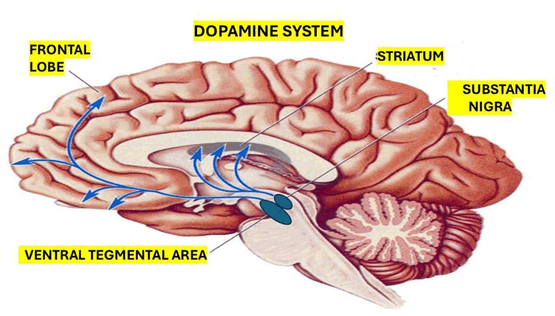
Figure 2: Shows the dopaminergic pathways
Parkinson’s Disease (PD) motor symptoms have been found to be primarily caused by disruption of dopaminergic pathways via the basal ganglia. But fascinating new research has shown how the circuitry of the basal ganglia plays a part in the development of depression in Parkinson’s disease. Research has demonstrated a correlation between elevated beta band activity in the globus pallidus and the intensity of depression symptoms [23]. Although this increased beta activity is usually associated with motor symptoms like bradykinesia and rigidity, it also suggests a type of “emotional rigidity,” which is marked by limited emotional expression, anhedonia, and decreased drive. This emphasizes how important it is for the basal ganglia to integrate motor, cognitive, and emotional functions. Therefore, rather than being limited to conventional mood-regulating pathways, it implies that the etiology of depression in Parkinson’s disease involves disruption in common motor-limbic networks.
The unique etiology of depression in Parkinson’s Disease (PD) is highlighted as a distinct neurobiological entity rather than just a comorbidity. Comparative PET imaging studies show that depression in Parkinson’s Disease (PD) and major depressive disorders share abnormalities involving the frontal and limbic regions, with extra involvement of motor and cognitive networks in depression linked to PD. This is demonstrated by changes in basal ganglia activity and hypometabolism in temporal areas [29].
Additionally, compared to limbic correlations, which are commonly seen in major depressive disorder or primary depression, the intensity of depressive symptoms in Parkinson’s Disease (PD) is more significantly correlated with sensorimotor and parietal cortex activity. However, we hypothesized that the comorbidity of depression and Parkinson’s Disease (PD) is closely linked to hippocampal dysfunction with damaged myelinated fibers of the fornix caused by injured oligodendrocytes. Figure 3 depicts the most essential components of that system.
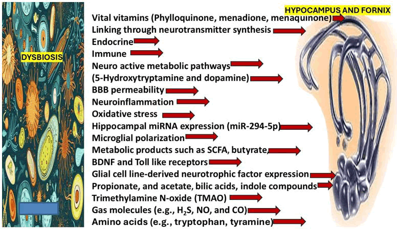
Figure 3: Shows a graphical representation of the microbiotas, the hippocampus, the fornix as a C-shaped bundle of white matter (part of the limbic system) located in the mesial aspect of both mesial temporal lobes just below the corpus callosum being the largest single pathway of the hippocampus and connecting it with other subcortical regions, diencephalon, basal forebrain and the thalamus. This graphic also shows the list of the most important elements involved in the pathogenesis of depression and NDD due to damage of the hippocampus and the fornix
Certain neuroimaging studies highlight the involvement of aberrant cortical-limbic networks in depression associated with Parkinson’s disease. Studies using functional imaging while at rest have revealed a decrease. Regional homogeneity in important regions related to emotional regulation, such as the prefrontal cortex, insula, and hippocampus [29]. Because these areas are essential for the integration of emotional and cognitive processes as well as the severity of depression, we think that limbic system dysfunction is a component of the mechanism of PD/depression.
Their functioning has been connected to symptoms. The idea that depression PD involves network-level dysfunction rather than only neurotransmitter imbalance is supported by reduced homogeneity within these networks, which indicates decreased coordination of cortico-limbic circuits.
All of these data point to the extensive effects of neurodegeneration beyond classical motor pathways, suggesting that depression in Parkinson’s disease arises from the convergence of motor, cognitive, and limbic network dysfunction [23].
The most important components of the limbic system are represented in Figure 4.
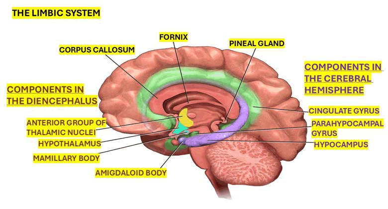
Figure 4: Shows the most important components of the limbic system
The disruption of cortico-limbic networks likely contributes to the emotional and cognitive aspects of depression seen in Parkinson’s disease, giving it a distinct presentation from depression in the general population, even though basal ganglia dysfunction may underlie motivational deficits as part of the non-motor symptoms.
Depression is closely associated with other non-motor symptoms of Parkinson’s Disease (PD), such as sleep difficulties, demonstrating its integration within the neurodegenerative process of the disease. According to research, sleep disturbances might contribute to cognitive dysfunction by causing depression, which in turn causes cognitive deterioration [30].
While REM sleep behavior disorder has both direct and indirect impacts on cognition, sleepiness appears to primarily affect cognitive decline through depressive pathways. These results point to overlapping symptom domains caused by common neurodegenerative processes affecting the brainstem, limbic, and cortical regions. This proof emphasizes that. Depression should not be seen as a separate illness since it is a key component of the intricate web of non-motor symptoms.
During the prodromal stage, there is more proof that depression in Parkinson’s disease is inherent. Other nonmotor symptoms may coexist with depressive symptoms long before the usual motor signs appear. This time pattern implies that rather than being a subsequent consequence of a chronic illness, depression may be an early clinical sign of the disease, arising from direct neurodegenerative effects. These findings have a biological basis due to the early involvement of limbic and monoaminergic pathways, which supports the idea that depression is a fundamental aspect of Parkinson’s disease.
Additional comments on depression in PD
Some investigators have reported confident evidence on a neurobiological substrate that supports that depression and anxiety can happen in the prodromal phase of Parkinson’s disease [31].
It has been documented that depression could be related to dysfunction in the prefrontal cortex and subcortical nuclei, the basal temporal limbic circuit, striatal-thalamicprefrontal cortex circuits, and brainstem monoamine and indolamine (i.e., dopamine, serotonin, and norepinephrine) systems.
Furthermore, genetic studies have found an association between genetic variants in SLC6A15, TPH2 [32] and BDNF [33] and PD’s depression, despite several investigations examining serotonin and dopamine transporter genes did not arrive to any confident conclusion while other researchers have found interrelationship between increased α-synuclein deposition in the substantia nigra, ventral tegmental area, and nucleus accumbens [34] neuronal loss in the posterolateral aspect of the par’s compacta substantia nigra [35] and changes in brain functional connectivity with EEG and functional MRI [36,37]. Small-vessel disease of the brain may also contribute to motor and non-motor symptoms in PD [38].
As mentioned before, there is novel evidence that changes in the microbiome or other gut changes might contribute to depression in Parkinson’s disease [39,40].
Depression may be considered a part of Neurodegenerative Disease (NDD) or a reaction to the onset of motor or nonmotor disability.
Brief comments on gut-brain axis and depression in Parkinson’s disease
Two years back, we documented the role of the Microbiota- Gut-Brain Axis (MGBA) in the pathogenesis of some neurological conditions when dysbiosis is present [41,42]. The clinical features of dysbiosis are shown in Figure 5.
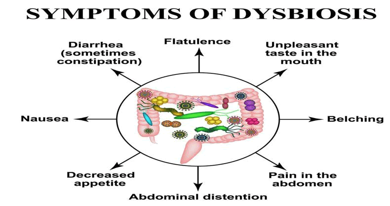
Figure 5: List of the most common clinical features of dysbiosis
Other writers have recently verified that the MGBA has a significant impact on the onset and course of NDDs. Through neurological, endocrine, immunological, and metabolic pathways, the gut microbiota and the central nervous system are connected by a complex bidirectional regulatory network known as the MGBA.
The Microbiota-Gut-Brain Axis (MGBA) significantly affects brain function through a number of pathways (symbiosis), including the immunological, neurological, endocrine, and metabolic systems (Figure 6). The degenerative processes of Parkinson’s disease are directly linked to their dysregulation. In particular, the Gut-Brain Axis (GBA), a process involving complex control of endocrine, immunological, and neurological signaling, allows the gut microbiota to communicate with the central nervous system in both directions [43].
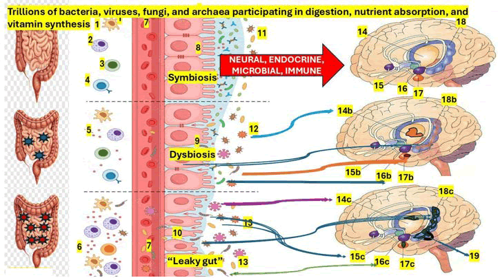
Figure 6: Graphical hypotheses of the pathophysiological effects on the limbic system by normal microbiotas and the target elements in the limbic system damaged by dysbiosis and “leaky gut”
Note: The pathogenesis of depression in PD involves several pathways, including synaptic dysfunction, protein homeostasis imbalance, pathological protein aggregation, mitochondrial dysfunction, neuroinflammation, ferroptosis, and interactions between genetic and environmental factors. 1) Dendritic cells, 2) Macrophages, 3) T-cell, 4) Cell, 5) Proinflammatory cytokines, 6) Inflammatory response, 7) Blood flow, 8) Symbiosis in normal intestinal epithelium, 9) Dysbiosis, 10) Leaky gut, 11) Normal microbiome, 12) Disbalance among Firmicutes and Bacteroidetes, 13) Pathogenic microbial growth, 14) Thalamus, 15) Hippocampus, 16) Hypothalamus, 17) Amygdala, 18) Hippocampus, 14b,15b,16b,17b, 18b) Moderate damaged areas of the limbic system by dysbiosis leading to PD/depression, 14c,15c,16c,17c,18c) Severely damaged areas of the limbic system by “leaky gut” leading to advanced PD and major depression, 19) Damaged pars compacta substantia nigra
It has been known for more than 20 years that the primary factor in the pathophysiology of Parkinson’s Disease (PD) is dysregulation of the MGBA, which modifies neurotransmitter synthesis, metabolite production, immune responses, and Blood-Brain Barrier (BBB) permeability, resulting in dysfunctional CNS homeostasis [44].
For instance, the GBA carries neuroactive metabolites (including dopamine and 5-hydroxytryptamine) and Short- Chain Fatty Acids (SCFAs) produced by the gut microbiota to the brain, where they control neuronal activity and encourage microglial polarization [45].
However, the gut, not the brain, produces more than 90% of the body’s serotonin (5-HT), a crucial neurotransmitter involved in mood regulation [46].
Additionally, a pathological progression of NDDs is caused by gut microbiota dysbiosis, or gut microbial imbalance, which results in oxidative stress and systemic neuroinflammation [47,48]. According to some writers, dysbiosis increases the BBB’s permeability and integrity by triggering aberrant immune responses and systemic chronic inflammation, which results in neurodegeneration and neuroinflammation (Figure 6) [41,42,49,50].
Currently, has been proven that depression and even most neurodegenerative pathologies are now linked to changes in the gut microbial community [51-53].
Histologically, the neuron cells of the ENS are organised into two principal ganglionated plexuses, the submucosal (Meissner’s) and the myenteric (Auerbach’s) plexuses [54].
We hypothesised that both plexuses, along with parasympathetic (VN) and sympathetic inputs, lead to a bidirectional link between the gut and brain, which is modulated via the MGBA, and it disbalance is the major component of the pathophysiology of PD/depression, as it has been suspected by other authors [55,56].
On the other hand, other investigators recommend VN stimulation to alleviate depression, anxiety, and epilepsy based on their role in modulating neural circuits related to mental health [57,58].
Apart from the role played by the VN in controlling inflammation, gut motility, and the secretion of digestive enzymes through its efferent fibres, it releases acetylcholine, 5-HT, dopamine, and Gamma-Aminobutyric Acid (GABA), and on top of that, its efferent fibres activate antiinflammatory response and downregulate pro-inflammatory cytokines to provide neuroinflammation [59].
As we represented in Figure 6 dysbiosis cause an impairment of the intestinal epithelial barrier enhancing permeability, a condition that in advance stage is named “leaky gut” [60] which disrupt the gut barrier function leading to the translocation of bacteria and/or their metabolic byproducts like lipopolysaccharides, from the lumen of the gut to the blood flow triggering a cascade of systemic inflammation including an upregulation of pro-inflammatory cytokines as graphically shown in Figure 6 as has been supported by other investigators [61,62].
Another consequence of the previously cited inflammatory state is disruption of the BBB, which reduces effectiveness as a protective filter, allowing peripheral immune cells to infiltrate the CNS. Particularly, in cases presenting PD with dysbiosis, migration of the CCR2+ monocytes from the gut into the brain is certainly contributing to progressive neurodegeneration [63].
Notwithstanding, elevated concentration of CCR2+ circulating monocytes has been reported in early PD cases with an associated cognitive decline [64].
Other investigators support the previous statement based on the mechanism by which gut dysbiosis and systemic inflammation may trigger the recruitment of CCR2+ monocytes into the brain, thereby driving neuroinflammation and neurodegeneration (PD), although they report that not all gut microbes exert similar effects on the host immune system [65,66].
While some microbiotas can drive the release of proinflammatory immune cells, such as Th17 cells, which are known to migrate to the CNS, leading to neuroinflammation, other beneficial microbiotas can foster the development of neuroregulatory T cells (NeuroTregs), which play a vital role in decreasing neuroinflammation [67].
As mentioned, several reports have consistently shown significant changes in the composition of gut microbiota in people with depression, with findings of reduced microbial diversity and different compositional shifts supporting the involvement of gut microbiota dysbiosis in the pathophysiology of depression [68-71].
Beyond histone modifications, gut microbiotas control post-transcriptional gene regulation through microRNAs (miRNAs) in the animal MDD model [72].
Another element involved in the pathogenesis of depression in PD is altered hippocampal miRNA expression, including miR-294-5p. Microbial colonisation restores miRNA levels, which can explain the influence of mood-related neurochemical pathways caused by Microbiota-mediated miRNA changes in the amygdala, the most important region for emotional regulation [73].
Therefore, the absence of gut microbes impaired amygdaladependent fear recall and altered the expression of genes linked to glutamatergic transmission, synaptic signalling, neurotrophic factors such as BDNF, and epigenetic regulation, supporting the idea that gut microbes modulate emotional behaviour through gene expression [74].
We hypothesised that the gut microbiota exerts neuroregulatory control through several epigenetic mechanisms, including miRNA-mediated gene silencing, chromatin remodelling, and modulation of RNA splicing, thereby making a significant contribution to the transcriptional control of key neural processes.
Based on our search of the medical literature, we hypothesised that α-synuclein aggregates may originate in the gut microbiota and spread from there to the brain, where neurons located on the posterolateral aspect of the pars compacta are selectively damaged, reducing their dopamine production. Other authors reported similar proposals but under different circumstances [75-79]. Therefore, the gut microbiome significantly influences brain chemistry by synthesising and metabolising various neuroactive molecules, including microbial metabolites and neurotransmitters such as serotonin, dopamine, and glutamate. On the other hand, SCFAs act as endogenous ligands for G protein-coupled receptors and, by inhibiting histone deacetylases, they regulate gene expression and neuroplasticity in the brain [80-82].
Other microbiotas metabolites, such as indole, secondary bile acids (deoxycholic acid and lithocholic acid), vital vitamins (menaquinone, phylloquinone, and menadione), amino acids (tyramine and tryptophan), LPS, long-chain fatty acids, and trimethylamine-N-oxide, can directly or indirectly promote the migration of peripheral immune cells to the brain leading to neuroinflammation, PD and other brain pathologies [83-86].
Inflammation also plays a crucial role in the mechanisms underlying the gut-brain axis in depression, which has been reported [87,88] in PD patients, reflecting decreased microbial diversity and ecological imbalance [89].
Despite these findings, some limitations should be considered when interpreting the results of this review including the small number of selected studies concerning to novel hypotheses of depression in PD, limited to publications from a narrow time frame within a single year, restricts the extent of available evidence and may not fully capture the complexity of the underlying mechanisms. In addition, most studies were cross-sectional in design, limiting the ability to establish causal relationships between neurobiological changes and depressive symptoms. Heterogeneity between studies is also influenced by differences in patient groups, assessment instruments, and imaging modalities. Additionally, patients on various pharmacological therapies and at different phases of the disease’s progression were included in numerous research, which may have an impact on both brain activity and clinical presentation. These elements emphasize the need for more extensive, long-term, and methodologically sound research to more clearly define the pathophysiology of depression in Parkinson’s disease.
Conclusion
In summary, dopaminergic dysfunction, network-level disturbances, dysbiosis, and more general neurodegenerative processes all seem to contribute to depression in Parkinson’s disease. Early detection and focused treatments are significantly impacted when depression is acknowledged as an inherent part of Parkinson’s Disease (PD) rather than a subsequent psychological reaction. Future therapeutic approaches to enhance mental and general illness outcomes may be guided by a better knowledge of these pathways.
To the best of our knowledge, this review is the first to concentrate on new facets of the pathophysiology of depression in Parkinson’s Disease (PD) and to present fresh theories on the impact of microbiota on the limbic system and other areas of the brain in Parkinsonian patients.
Acknowledgment
To thanks to Prof Thozama Dubula for his unconditional support.
Ethics Statement
This review does not require ethical approval.
Patient Privacy
All patient-identifying information has been removed to ensure anonymity.
Conflicts of Interest
Authors of this review report there is not conflicts of interest.
References
- L. Kondrataviciute, M. Kapadia, H. Chau, C. Tan, P. Ou, et al. Characterization of motor and non-motor features associated with bilateral nigral degeneration due to A53T alpha-synuclein in female rats, Sci Rep, 16(2026):5462.
[Crossref] [Google Scholar] [PubMed]
- A. Schrag, Quality of life and depression in Parkinson's disease, J Neurol Sci, 248(2006):151–157.
[Crossref] [Google Scholar] [PubMed]
- R. Chikatimalla, T. Dasaradhan, J. Koneti, S.P. Cherukuri, R. Kalluru, et al. Depression in Parkinson's disease: A narrative review, Cureus, 14(2022):e27750.
[Crossref] [Google Scholar] [PubMed]
- J. Zhao, H. Jia, P. Ma, D. Zhu, Y. Fang, Multidimensional mechanisms of anxiety and depression in Parkinson's disease: Integrating neuroimaging, neurocircuits, and molecular pathways, Pharmacol Res, 212(2025):107717.
[Crossref] [Google Scholar] [PubMed]
- M. H. Ahmad, M.A. Rizvi, M. Ali, A.C. Mondal, Neurobiology of depression in Parkinson's disease: Insights into epidemiology, molecular mechanisms and treatment strategies, Ageing Res Rev, 84(2023):101840.
[Crossref] [Google Scholar] [PubMed]
- J. G. Goldman, R. Postuma, Premotor and nonmotor features of Parkinson's disease, Curr Opin Neurol, 27(2014):434–441.
[Crossref] [Google Scholar] [PubMed]
- N. Zhao, Y. Yang, L. Zhang, Q. Zhang, L. Balbuena, et al. Quality of life in Parkinson's disease: A systematic review and meta-analysis of comparative studies, CNS Neurosci Ther, 27(2021):270–279.
[Crossref] [Google Scholar] [PubMed]
- American Psychiatric Association, Diagnostic and statistical manual of mental disorders: DSM-5-TR. Washington, DC: American Psychiatric Association Publishing; (2022).
- K. A. Jellinger, The pathobiological basis of depression in Parkinson disease: Challenges and outlooks, J Neural Transm, 129(2022):1397–1418.
[Crossref] [Google Scholar] [PubMed]
- D. Weintraub, D. Aarsland, K. R. Chaudhuri, R. D. Dobkin, A. F. G. Leentjens, et al. The neuropsychiatry of Parkinson's disease: Advances and challenges, Lancet Neurol, 21(2022):89–102.
[Crossref] [Google Scholar] [PubMed]
- R. Kim, J. H. Shin, S. Park, H. J. Kim, B. Jeon, Longitudinal evolution of non-motor symptoms according to age at onset in early Parkinson's disease, J Neurol Sci, 418(2020):117157.
[Crossref] [Google Scholar] [PubMed]
- B. Ellul, A. McNamara, S. Laurenz, I. Baetu, M. Jenkinson, et al. Predicting cognition and affective changes in newly diagnosed Parkinson's disease through longitudinal data-driven clustering, J Geriatr Psychiatry Neurol, 39(2025):310–326.
[Crossref] [Google Scholar] [PubMed]
- E. Fiorenzato, R. Biundo, D. Cecchin, A.C. Frigo, J. Kim, et al. Brain amyloid contribution to cognitive dysfunction in early-stage Parkinson's disease: The PPMI dataset, J Alzheimers Dis, 66(2018):229–237.
[Crossref] [Google Scholar] [PubMed]
- R. M. Meade, D. P. Fairlie, J. M. Mason, Alpha-synuclein structure and Parkinson's disease: Lessons and emerging principles, Mol Neurodegener, 14(2019):29.
[Crossref] [Google Scholar] [PubMed]
- D. R. Howlett, D. Whitfield, M. Johnson, J. Attems, J.T. O'Brien, et al. Regional multiple pathology scores are associated with cognitive decline in Lewy body dementias, Brain Pathol, 25(2015):401–408.
[Crossref] [Google Scholar] [PubMed]
- F. G. Leentjens, R. Lousberg, F. R. J. Verhey, Markers for depression in Parkinson's disease, Acta Psychiatr Scand, 106(2002):196–201.
[Crossref] [Google Scholar] [PubMed]
- S. C. Gu, J. Zhou, C. X. Yuan, Q. Ye, Personalized prediction of depression in patients with newly diagnosed Parkinson's disease: A prospective cohort study, J Affect Disord, 268(2020):118–126.
[Crossref] [Google Scholar] [PubMed]
- E. Candel-Parra, M. P. Córcoles-Jiménez, V. Delicado-Useros, M. C. Ruiz-Grao, A. Hernández-Martínez, et al. Predictive model of quality of life in patients with Parkinson's disease, Int J Environ Res Public Health, 19(2022):672.
[Crossref] [Google Scholar] [PubMed]
- R. Rajmohan, P. H. Reddy, Amyloid-beta and phosphorylated tau accumulations cause abnormalities at synapses of Alzheimer's disease neurons, J Alzheimers Dis, 57(2017):975–999.
[Crossref] [Google Scholar] [PubMed]
- P. Lei, S. Ayton, D. I. Finkelstein, P. A. Adlard, C. L. Masters, et al. Tau protein: Relevance to Parkinson's disease, Int J Biochem Cell Biol, 42(2010):1775–1778.
[Crossref] [Google Scholar] [PubMed]
- G. Alves, K. Bronnick, D. Aarsland, K. Blennow, H. Zetterberg, et al. CSF amyloid-beta and tau proteins, and cognitive performance, in early and untreated Parkinson's disease: The Norwegian ParkWest study, J Neurol Neurosurg Psychiatry, 81(2010):1080–1086.
[Crossref] [Google Scholar] [PubMed]
- J. Yang, X. Song, S. Yan, Q. Li, W. Yang, The gut microbiota influences neurodegenerative diseases through the gut-brain axis: Molecular mechanisms and effects on immune function, Front Immunol, 16(2026):1739329.
[Crossref] [Google Scholar] [PubMed]
- K. A. Johnson, P. B. Coutinho, L. E. Kenney, J. K. Wong, J. D. Hilliard, et al. Pallidal beta power is associated with depression in Parkinson's disease, NPJ Parkinsons Dis, 12(2026):50.
[Crossref] [Google Scholar] [PubMed]
- A. Schrag, L. Horsfall, K. Walters, A. Noyce, I. Petersen, Prediagnostic presentations of Parkinson's disease in primary care: A case-control study, Lancet Neurol, 14(2015):57–64.
[Crossref] [Google Scholar] [PubMed]
- A. L. A. J. Hommel, M. J. Meinders, S. Lorenzl, R. Dodel, M. Coelho, et al. The prevalence and determinants of neuropsychiatric symptoms in late-stage parkinsonism, Mov Disord Clin Pract (Hoboken), 7(2020):531–542.
[Crossref] [Google Scholar] [PubMed]
- R. Ou, J. Lin, K. Liu, Z. Jiang, Q. Wei, et al. Evolution of apathy in early Parkinson's disease: A 4-years prospective cohort study, Front Aging Neurosci, 12(2021):620762.
[Crossref] [Google Scholar] [PubMed]
- B. M. Scott, R. S. Eisinger, M. R. Burns, J. Lopes, M.S. Okun, et al. Co-occurrence of apathy and impulse control disorders in Parkinson disease, Neurology, 95(2020):e2769–e2780.
[Crossref] [Google Scholar] [PubMed]
- D. Weintraub, M. Caspell-Garcia, T. Simuni, H.R. Cho, C.S. Coffey, et al. Neuropsychiatric symptoms and cognitive abilities over the initial quinquennium of Parkinson disease, Ann Clin Transl Neurol, 7(2020):449–461.
[Crossref] [Google Scholar] [PubMed]
- M. Liu, X. Xie, Y. Liu, H. Feng, X. Wang, et al. Comparison of clinical features and cerebral glucose metabolism between patients with major depressive disorder and Parkinson's with depression, BMC Psychiatry, 26(2026).
[Crossref] [Google Scholar] [PubMed]
- H. Zhao, Y. Sun, Q. Wang, S. Li, Z. Zhao, Depression mediated sleep disturbances and cognitive decline in Parkinson's disease patient, Front Neurosci, 20(2026):1722979.
- A. Schrag, L. Horsfall, K. Walters, A. Noyce, I. Petersen, Prediagnostic presentations of Parkinson's disease in primary care: A case-control study, Lancet Neurol, 14(2015):57–64.
[Crossref] [Google Scholar] [PubMed]
- J. Zheng, X. Yang, Q. Zhao, S. Tian, H. Huang, et al. Association between gene polymorphism and depression in Parkinson's disease: A case-control study, J Neurol Sci, 375(2017):231–234.
[Crossref] [Google Scholar] [PubMed]
- M. Ramezani, J. A. Ruskey, K. Martens, M. Kibreab, Z. Javer, et al. Association between BDNF Val66Met polymorphism and mild behavioral impairment in patients with Parkinson's disease, Front Neurol, 11(2021):587992.
[Crossref] [Google Scholar] [PubMed]
- L. Patterson, S. P. Rushton, J. Attems, A. J. Thomas, C. M. Morris, Degeneration of dopaminergic circuitry influences depressive symptoms in Lewy body disorders, Brain Pathol, 29(2019):544–557.
[Crossref] [Google Scholar] [PubMed]
- L. Saari, L. Heiskanen, M. Gardberg, V. Kaasinen, Depression and nigral neuron density in Lewy body spectrum diseases, Ann Neurol, 89(2021):1046–1050.
[Crossref] [Google Scholar] [PubMed]
- K. K. Iyer, T. R. Au, A. J. Angwin, D. A. Copland, N. N. Dissanayaka, Theta and gamma connectivity is linked with affective and cognitive symptoms in Parkinson's disease, J Affect Disord, 277(2020):875–884.
[Crossref] [Google Scholar] [PubMed]
- H. Liao, S. Cai, Q. Shen, J. Fan, T. Wang, et al. Networks are associated with depression in patients with Parkinson's disease: A resting-state imaging study, Front Neurosci, 14(2021):573538.
[Crossref] [Google Scholar] [PubMed]
- H. Chen, H. Wan, M. Zhang, G. Liu, X. Wang, et al. Cerebral small vessel disease may worsen motor function, cognition, and mood in Parkinson's disease, Parkinsonism Relat Disord, 83(2021):86–92.
[Crossref] [Google Scholar] [PubMed]
- O. I. Olasunkanmi, L. Zheng, P. Zheng, Gut brain axis in health and brain disease, Chin Med J (Engl), 139(2026):799–827.
- C. Girges, N. Vijiaratnam, A. King, G. Auld, R. McComish, et al. Mild-to-moderate depressive symptoms impact on self-reported outcome measures in clinical trials for neurodegenerative diseases, Clin Trials, 23(2026):54–64.
[Crossref] [Google Scholar] [PubMed]
- L. F. I. Valdes, H. F. Sibat, Comorbidity of alcohol use disorder and neurocysticercosis aggravated by dysbiosis, J Drug Alcohol Res, 13(2024):236417.
- L. F. I. Valdes, H. F. Sibat, Impact of gut dysbiosis on neuromyelitis optica spectrum disorder: Implications for alcohol and substance use, J Drug Alcohol Res, 13(2024):236418.
- J. Yang, X. Song, S. Yan, Q. Li, W. Yang, The gut microbiota influences neurodegenerative diseases through the gut-brain axis: Molecular mechanisms and effects on immune function, Front Immunol, 16(2026):1739329.
[Crossref] [Google Scholar] [PubMed]
- R. H. Howland, Vagus nerve stimulation, Curr Behav Neurosci Rep, 1(2014):64–73.
[Crossref] [Google Scholar] [PubMed]
- K. G. Margolis, J. F. Cryan, E. A. Mayer, The microbiota-gut-brain axis: From motility to mood, Gastroenterology, 160(2021):1486–1501.
[Crossref] [Google Scholar] [PubMed]
- N. Akram, Z. Faisal, R. Irfan, Y. A. Shah, S. A. Batool, et al. Exploring the serotonin-probiotics-gut health axis: A review of current evidence and potential mechanisms, Food Sci Nutr, 12(2024):694–706.
[Crossref] [Google Scholar] [PubMed]
- S. Hamamah, A. Aghazarian, A. Nazaryan, A. Hajnal, M. Covasa, Role of microbiota-gut-brain axis in regulating dopaminergic signalling, Biomedicines, 10(2022):436.
[Crossref] [Google Scholar] [PubMed]
- C. Tan, Q. Yan, Y. Ma, J. Fang, Y. Yang, Recognizing the role of the vagus nerve in depression from microbiota-gut brain axis, Front Neurol, 13(2022):1015175.
[Crossref] [Google Scholar] [PubMed]
- L. Y. Kamel, W. Xiong, B. M. Gott, A. Kumar, C. R. Conway, Vagus nerve stimulation: An update on a novel treatment for treatment-resistant depression, J Neurol Sci, 434(2022):120171.
[Crossref] [Google Scholar] [PubMed]
- V. A. Pavlov, K. J. Tracey, The cholinergic anti-inflammatory pathway, Brain Behav Immun, 19(2005):493–499.
[Crossref] [Google Scholar] [PubMed]
- X. Hu, Y. Li, J. Wu, H. Zhang, Y. Huang, et al. Changes of gut microbiota reflect the severity of major depressive disorder: A cross-sectional study, Transl Psychiatry, 13(2023):137.
[Crossref] [Google Scholar] [PubMed]
- P. Zheng, J. Yang, Y. Li, J. Wu, W. Liang, et al. Gut microbial signatures can discriminate unipolar from bipolar depression, Adv Sci (Weinh), 7(2020):1902862.
[Crossref] [Google Scholar] [PubMed]
- P. Zheng, B. Zeng, C. Zhou, M. Liu, Z. Fang, et al. Gut microbiome remodelling induces depressive-like behaviours through a pathway mediated by the host's metabolism, Mol Psychiatry, 21(2016):786–796.
[Crossref] [Google Scholar] [PubMed]
- Z. H. Geng, Y. Zhu, Q. L. Li, C. Zhao, P. H. Zhou, Enteric nervous system: The bridge between the gut microbiota and neurological disorders, Front Aging Neurosci, 14(2022):810483.
[Crossref] [Google Scholar] [PubMed]
- S. Breit, A. Kupferberg, G. Rogler, G. Hasler, Vagus nerve as modulator of the brain-gut axis in psychiatric and inflammatory disorders, Front Psychiatry, 9(2018):44.
[Crossref] [Google Scholar] [PubMed]
- K. G. Margolis, J. F. Cryan, E. A. Mayer, The microbiota-gut-brain axis: From motility to mood, Gastroenterology, 160(2021):1486–1501.
[Crossref] [Google Scholar] [PubMed]
- C. Tan, Q. Yan, Y. Ma, J. Fang, Y. Yang, Recognizing the role of the vagus nerve in depression from microbiota-gut brain axis, Front Neurol, 13(2022):1015175.
[Crossref] [Google Scholar] [PubMed]
- L. Y. Kamel, W. Xiong, B. M. Gott, A. Kumar, C. R. Conway, Vagus nerve stimulation: An update on a novel treatment for treatment-resistant depression, J Neurol Sci, 434(2022):120171.
[Crossref] [Google Scholar] [PubMed]
- V. A. Pavlov, K. J. Tracey, The cholinergic anti-inflammatory pathway, Brain Behav Immun, 19(2005):493–499.
[Crossref] [Google Scholar] [PubMed]
- Y. Kinashi, K. Hase, Partners in leaky gut syndrome: Intestinal dysbiosis and autoimmunity, Front Immunol, 12(2021):673708.
[Crossref] [Google Scholar] [PubMed]
- L. An, U. Wirth, D. Koch, M. Schirren, M. Drefs, et al. The role of gut-derived lipopolysaccharides and the intestinal barrier in fatty liver diseases, J Gastrointest Surg, 26(2022):671–683.
[Crossref] [Google Scholar] [PubMed]
- J. Camberos-Barraza, A. M. Guadron-Llanos, A. K. De la Herran-Arita, The gut microbiome-neuroglia axis: Implications for brain health, inflammation, and disease, Neuroglia, 5(2024):254–273.
- A. S. Harms, A. D. Thome, Z. Yan, A. M. Schonhoff, G. P. Williams, et al. Peripheral monocyte entry is required for alpha-synuclein induced inflammation and neurodegeneration in a model of Parkinson disease, Exp Neurol, 300(2018):179–187.
[Crossref] [Google Scholar] [PubMed]
- S. K. Nissen, K. Farmen, M. Carstensen, C. Schulte, D. Goldeck, et al. Changes in CD163+, CD11b+, and CCR2+ peripheral monocytes relate to Parkinson’s disease and cognition, Brain Behav Immun, 101(2022):182–193.
[Crossref] [Google Scholar] [PubMed]
- A. K. DeGruttola, D. Low, A. Mizoguchi, E. Mizoguchi, Current understanding of dysbiosis in disease in human and animal models, Inflamm Bowel Dis, 22(2016):1137–1150.
[Crossref] [Google Scholar] [PubMed]
- D. Zheng, T. Liwinski, E. Elinav, Interaction between microbiota and immunity in health and disease, Cell Res, 30(2020):492–506.
[Crossref] [Google Scholar] [PubMed]
- Y. Shi, B. Wei, L. Li, B. Wang, M. Sun, Th17 cells and inflammation in neurological disorders: Possible mechanisms of action, Front Immunol, 13(2022):932152.
[Crossref] [Google Scholar] [PubMed]
- Y. Cheng, Z. Zhu, Z. Yang, X. Liu, X. Qian, et al. Alterations in fecal microbiota composition and cytokine expression profiles in adolescents with depression: A case-control study, Sci Rep, 15(2025):12177.
[Crossref] [Google Scholar] [PubMed]
- A. Hernandez-Cacho, J. F. Garcia-Gavilan, A. Atzeni, P. Konstanti, C. Belzer, et al. Multi-omics approach identifies gut microbiota variations associated with depression, NPJ Biofilms Microbiomes, 11(2025):68.
[Crossref] [Google Scholar] [PubMed]
- M. Meng, Gut microbiota variations in depression and anxiety: A systematic review, BMC Psychiatry, 25(2025):443.
[Crossref] [Google Scholar] [PubMed]
- X. Hu, Y. Li, J. Wu, H. Zhang, Y. Huang, et al. Changes of gut microbiota reflect the severity of major depressive disorder: A cross-sectional study, Transl Psychiatry, 13(2023):137.
[Crossref] [Google Scholar] [PubMed]
- G. M. Moloney, O. F. O’Leary, E. Salvo-Romero, L. Desbonnet, F. Shanahan, et al. Microbial regulation of hippocampal miRNA expression: Implications for transcription of kynurenine pathway enzymes, Behav Brain Res, 334(2017):50–54.
[Crossref] [Google Scholar] [PubMed]
- R. M. Stilling, G. M. Moloney, F. J. Ryan, A. E. Hoban, F. Shanahan, et al. Social interaction-induced activation of RNA splicing in the amygdala of microbiome-deficient mice, eLife, 7(2018).
[Crossref] [Google Scholar] [PubMed]
- A. E. Hoban, R. M. Stilling, G. Moloney, F. Shanahan, T. G. Dinan, et al. The microbiome regulates amygdala-dependent fear recall, Mol Psychiatry, 23(2018):1134–1144.
[Crossref] [Google Scholar] [PubMed]
- L. Klingelhoefer, H. Reichmann, Pathogenesis of Parkinson disease-the gut-brain axis and environmental factors, Nat Rev Neurol, 11(2015):625–636.
[Crossref] [Google Scholar] [PubMed]
- N. Uemura, H. Yagi, M. T. Uemura, Y. Hatanaka, H. Yamakado, et al. Inoculation of α-synuclein preformed fibrils into the mouse gastrointestinal tract induces Lewy body-like aggregates in the brainstem via the vagus nerve, Mol Neurodegener, 13(2018):21.
[Crossref] [Google Scholar] [PubMed]
- C. Challis, A. Hori, T. R. Sampson, B. B. Yoo, R. C. Challis, et al. Gut-seeded α-synuclein fibrils promote gut dysfunction and brain pathology specifically in aged mice, Nat Neurosci, 23(2020):327–336.
[Crossref] [Google Scholar] [PubMed]
- D. Pietrucci, R. Cerroni, V. Unida, A. Farcomeni, M. Pierantozzi, et al. Dysbiosis of gut microbiota in a selected population of Parkinson’s patients, Parkinsonism Relat Disord, 65(2019):124–130.
[Crossref] [Google Scholar] [PubMed]
- T. R. Sampson, C. Challis, N. Jain, A. Moiseyenko, M. S. Ladinsky, et al. A gut bacterial amyloid promotes α-synuclein aggregation and motor impairment in mice, eLife, 9(2020).
[Crossref] [Google Scholar] [PubMed]
- B. Dalile, L. van Oudenhove, B. Vervliet, K. Verbeke, The role of short-chain fatty acids in microbiota gut brain communication, Nat Rev Gastroenterol Hepatol, 16(2019):461–478.
[Crossref] [Google Scholar] [PubMed]
- M.W. Bourassa, I. Alim, S.J. Bultman, R.R. Ratan, Butyrate, neuroepigenetics and the gut microbiome: Can a high fiber diet improve brain health?, Neurosci Lett, 625(2016):56–63.
[Crossref] [Google Scholar] [PubMed]
- S.M. Matt, J.M. Allen, M.A. Lawson, L.J. Mailing, J.A. Woods, et al. Butyrate and dietary soluble fiber improve neuroinflammation associated with aging in mice, Front Immunol, 9(2018):1832.
[Crossref] [Google Scholar] [PubMed]
- T.R. Sampson, J.W. Debelius, T. Thron, S. Janssen, G.G. Shastri, et al. Gut microbiota regulate motor deficits and neuroinflammation in a model of Parkinson disease, Cell, 167(2016):1469–1480.e1412.
- J.S. Loh, W.Q. Mak, L.K.S. Tan, C.X. Ng, H.H. Chan, et al. Microbiota-gut-brain axis and its therapeutic applications in neurodegenerative diseases, Signal Transduct Target Ther, 9(2024):37.
[Crossref] [Google Scholar] [PubMed]
- P. Li, B.A. Killinger, E. Ensink, I. Beddows, A. Yilmaz, et al. Gut microbiota dysbiosis is associated with elevated bile acids in Parkinson’s disease, Metabolites, 11(2021):29.
[Crossref] [Google Scholar] [PubMed]
- F.L. da Silva, E. Coelho Cerqueira, M.S. de Freitas, D.L. Gonçalves, L.T. Costa, et al. Vitamins K interact with N-terminus α-synuclein and modulate the protein fibrillization in vitro: Exploring the interaction between quinones and α-synuclein, Neurochem Int, 62(2013):103–112.
[Crossref] [Google Scholar] [PubMed]
- E.F. Osimo, T. Pillinger, I.M. Rodriguez, G.M. Khandaker, C.M. Pariante, et al. Inflammatory markers in depression: A meta-analysis of mean differences and variability in 5,166 patients and 5,083 controls, Brain Behav Immun, 87(2020):901–909.
[Crossref] [Google Scholar] [PubMed]
- R. Shi, X. Gwee, D.Q.L. Chua, C.T.Y. Tan, K.B. Yap, et al. Inflammatory markers and incident depression: Evidence in a population-based prospective study, Psychoneuroendocrinology, 142(2022):105806.
[Crossref] [Google Scholar] [PubMed]
- T. Ji, H. Huang, J. Liu, T. Peng, X. Zhou, et al. Leveraging sequence-based faecal microbial community survey data to identify alterations in gut microbiota among patients with Parkinson’s disease, Eur J Neurosci, 53(2021):687–696.
[Crossref] [Google Scholar] [PubMed]
Copyright: © 2026 Saumya Mathew, et al. This is an open access article distributed under the terms of the Creative Commons Attribution License, which permits unrestricted use, distribution and reproduction in any medium, provided the original work is properly cited.