
Research Article: Journal of Drug and Alcohol Research (2026) Volume 15, Issue 6
New Hypotheses on Midbrain Stroke Secondary to HIV Vasculitis and Drug Management
Lourdes de Fatima Ibanez Valdes* and Humberto Foyaca SibatLourdes de Fatima Ibanez Valdes, Department of Internal Medicine and Therapeutics, Nelson Mandela Academic Hospital, Walter Sisulu University, South Africa, Email: humbertofoyacasibat@gmail.co
Received: 13-Mar-2026, Manuscript No. JDAR-26-192570; Editor assigned: 16-Mar-2026, Pre QC No. JDAR-26-192570 (PQ); Reviewed: 30-Mar-2026, QC No. JDAR-26-192570; Revised: 04-Jun-2026, Manuscript No. JDAR-26-192570 (R); Published: 11-Jun-2026, DOI: 10.4303/JDAR/236518
Abstract
Introduction: Midbrain ischemic stroke represents a condition mainly caused by an occlusion of the Posterior Cerebral Artery (PCA) branches, which are paired terminal branches of the basilar artery in the circle of Willis. PCA provides supply to most parts of the occipital cortex, the inferior and medial temporal cortices, the thalamus, and rostral midbrain. In the pre-ART era, Patients Living with HIV (PLWH) had a high risk of ischemic stroke compared to those without HIV, and the strokes occurred mostly in those with advanced AIDS, complicated with secondary infections such as fungal meningitis, toxoplasmosis encephalitis, tuberculous meningitis and neurosyphilis or those with coagulopathies or vasculitis.
The main aid of this review is to report a case presenting a midbrain stroke due to HIV vasculitis and to bring more light into the pathogenesis of this condition regarding its pathogenesis on vascular occlusion and reperfusion.
Methods: A large search of the articles published in SCOPUS, and MEDLINE to identify manuscripts related to midbrain ischemic stroke related to HIV vasculitis. From 01st, January 1990 to 30th, January 2026, we comprehensively review the medical literature and selected the following issues: “HIV vasculitis” OR “midbrain stroke” OR “Weber syndrome” OR “HIV-atherosclerosis” OR “pathophysiology of occlusion/reperfusion”, OR “pathophysiologic of midbrain stroke” OR “drug management of midbrain stroke”.
Results: After searching medical publications, 407 articles were retrieved. We removed 291 duplicated titles and abstracts; 116 manuscripts are chosen. After assessing the inclusion/exclusion related to HIV-vasculitis midbrain stroke and pathogenesis of reperfusion criteria all publications were removed; finally, cero articles investigated the pathogenesis of occlusion and reperfusion of ischemic midbrain stroke secondary to HIV vasculitis.
Conclusions: Based on our comprehensive review of the medical literature, we concluded that Posterior Cerebral Artery (PCA) midbrain stroke represents a smaller proportion of IS, often results in significant neurological deficits and poses distinctive therapeutic challenges.
As far as we know, this is the first study that graphically represent the elements involved in the pathogenesis of midbrain stroke secondary to HIV vasculitis and its mechanism of reperfusion at the rostral midbrain.
Keywords
Introduction
Midbrain ischemic stroke represents a condition mainly caused by an occlusion of the Posterior Cerebral Artery (PCA) branches, which are paired terminal branches of the basilar artery in the circle of Willis. PCA provides supply to most parts of the occipital cortex, the inferior and medial temporal cortices, the thalamus, and rostral midbrain. PCA strokes comprise 6–9% of all IS [1].
Most PCA obstructions are caused by embolic material, mainly from a cardiac source (24%), an arterial source (14%), or large-artery atherosclerosis (32%) [2].
Other less common aetiologies include arterial dissection, vasculitis, fibromuscular dysplasia, and moyamoya disease [3]. On the other hand, the Best Medical Treatment (BMT) for midbrain stroke traditionally includes antiplatelet agents, anticoagulants, statins, and supportive care [3].
Ipsilateral third nerve palsy and contralateral hemiparesis are the hallmarks of Weber’s syndrome. However, it’s an uncommon sign of brainstem stroke compared with supratentorial region stroke. Midbrain infarction is the commonest cause of Weber’s syndrome. The perforating branches of the basilar bifurcation or occlusion of the posterior cerebral artery commonly cause Ischemic Stroke (IS). Other investigators reported that isolated midbrain infarction accounts for less than 1% of posterior circulation cases [4,5].
Only a few case reports have documented the classic figure of Weber’s syndrome [6-8].
Human Immunodeficiency Virus (HIV) infection remains a significant global health concern, with millions affected worldwide [9].
HIV infection can present with a variety of nonspecific symptoms, often delaying diagnosis. Neurological and autoimmune complications have been associated with HIV-induced immune dysregulation from the direct action of the virus on the central nervous system. Although HIVassociated vasculitis is rare in many countries, in our region it is quite common; its recognition is crucial due to its potential impact on disease outcome and drug management. The confirmation of HIV-associated vasculitis requires detailed clinical assessment and imaging studies. ART remains the cornerstone of treatment, controlling viral replication, reducing immune dysregulation, and improving systemic inflammation. Early diagnosis and prompt initiation of ART play a critical role in preventing complications and ensuring a favourable long-term prognosis [10].
The first phase of HIV infection classically presents within two to four weeks after exposure, during which many individuals experience Acute Retroviral Syndrome (ARS). This period is typically characterised by nonspecific symptoms such as pharyngitis, rash, fever, lymphadenopathy, myalgia, and headache [11].
It is crucial to note that while these symptoms are generally self-limited, they are associated with high viral replication and a marked increase in HIV transmission risk. Nevertheless, this acute phase can present a transient decline in CD4+ T-cell counts, which sometimes recovers fully after the initial immune response [3].
Soon after the acute phase, people can experience a prolonged asymptomatic period, known as clinical latency, which can last several years. There is growing evidence linking HIV infection with exacerbations of autoimmune disorders and vasculitis, which can further obscure diagnosis [12]. The interplay between HIV infection, autoimmune conditions, and vasculitis requires heightened clinical suspicion, as it may significantly impact the course and management of patients [11].
The global incidence of ischemic stroke among patients with HIV is increasing gradually. Schaefer et al., conducted a retrospective, observational study of HIV-positive patients treated between 2012 and 2018, and they found that 0.7% were newly or previously diagnosed as HIV-positive. The same authors reported that IS were caused by large-vessel disease (37.5%), small-vessel disease (20.8%), cryptogenic embolism (20.8%), vasculitis (16.7%), and cardioembolism (4.2%). They also reported that large-vessel disease-related strokes were more often located in the posterior circulation (77.8%), the HIV-related cerebral vasculitis was associated with high mortality (75%), and the incidence of IS in HIVpositive peoples appears to be higher than in the general population, and the age at onset appears to be younger [12] as was previously confirmed by other investigators [13].
Chronic infections as well as HIV medication itself might increase the risk of stroke based on the pathophysiological explanations, which include atherosclerosis fostered through chronic inflammation or dyslipidaemia due to antiretroviral medication [14].
The assumption of a specifically HIV-associated vasculitis and its differentiation from common atherosclerosis and vasculitis have been reported before [15].
In 1986, Ischemic Stroke (IS) was first reported in patients with HIV infection by Anders and colleagues [16].
The incidence of IS was reported to be higher in HIVinfected children and young adults, with a lack of traditional risk factors [17,18].
In the Pre-ART era, Patients Living with HIV (PLWH) had a high risk of ischemic stroke compared to those without HIV, and the strokes occurred mostly in those with advanced AIDS, complicated with secondary infections such as fungal meningitis, toxoplasmosis encephalitis, tuberculous meningitis and neurosyphilis or those with coagulopathies or vasculitis [19-21].
On the other hand, Cole et al., reported that in the pre-ART population, the incidence of ischemic stroke was nine times higher in AIDS patients than in their control group [22].
In the post-ART era, other investigations confirmed an increased risk of IS in PLWH independent of age and traditional vascular risk factors [22].
Genetic factors also highlighted the increased risk of IS in HIV infected patients. Some investigations have shown an increased risk of IS in African Americans in ART-treated PLWH compared with other race/ethnic groups and a greater risk of stroke in women and younger people in the US population [23,24].
Nevertheless, it has been proven that HIV remain quiescent in many cells of the body, including those of the CNS [25]. Therefore, viral reservoirs are being confirmed in microglia, astrocytes, perivascular macrophages, and pericytes [26,27].
Although Retroviral Therapy (ART) has moved the outcome of HIV-positive infection from a fatal condition to a chronic disease. As previously cited, treated HIV patients have a high prevalence of HIV associated comorbidities, including ischemic stroke. Despite the aetiology of stroke being HIVrelated remaining unknown, several factors such as vascular abnormalities, opportunistic infections, atherosclerosis and diabetes can contribute to the pathogenesis of stroke. In addition, chronic administration of ART contributes to the increased risk of stroke in HIV infected patients. Some investigators reported that murine model of ischemic stroke have HIV infection worsens stroke outcome, increases blood brain barrier permeability and increases neuroinflammation and residual HIV viral proteins, such as trans-activator of transcription, glycoprotein 120 and negative regulatory factor are involved in its pathogenesis [28].
The main aid of this review is to report a case presenting a midbrain stroke due to HIV vasculitis and to bring more light into the pathogenesis of this condition regarding its pathogenesis on vascular occlusion and reperfusion.
Materials and Methods
A large search of the articles published in SCOPUS, and MEDLINE to identify manuscripts related to midbrain ischemic stroke related to HIV vasculitis.
From 01st, January 1990 to 30th, January 2026, we comprehensively review the medical literature and selected the following issues: “HIV vasculitis” OR “midbrain stroke” OR “Weber syndrome” OR “HIV-atherosclerosis” OR “pathophysiology of occlusion/reperfusion”, OR “pathophysiologic of midbrain stroke” OR “drug management of midbrain stroke”.
Results and Discussion
Literature search
After searching medical publications, 407 articles were retrieved. We removed 291 duplicated titles and abstracts; 116 manuscripts are chosen. After assessing the inclusion/exclusion related to HIV-vasculitis midbrain stroke and pathogenesis of reperfusion criteria all publications were removed; finally, cero articles investigated the pathogenesis of occlusion and reperfusion of ischemic midbrain stroke secondary to HIV vasculitis.
Mrs ZM is a 37-year-old female, RVD-reactive, and on ART since December last year. She complained of difficulty seeing in both eyes, which started eight months ago and progressively worsened. It was alleviated by reading glasses, with difficulty seeing distant objects still visible; if she was not wearing them, she closed one eye to see better. Worsened when outside exposed in the sun and was associated with itchy, painful eyes. Weakness in the right arm and leg began 7 months ago; she noticed that when carrying things, they would fall and that people noticed she was limping. Seven months ago, she attended a traditional healer and got better, but the weakness suddenly struck heavily two months later, and she decided to come to the hospital. The double vision started six months ago; it has improved since she attended the clinic.
Physical examination: Sitting on the bed, reading glasses next to the patient; ptosis noted. The face is dry with hypopigmented patches.
General examination: Unremarkable. VITALS, BP: 130/93, PR: 76 bpm, T: 36.8°C, RR: 18 bpm.
Neurological examination: The patient is alert and welloriented to time, place, and person. GCS: 15/15, CN1: Intact, CNII: Visual fields clear in all fields, fundoscopy normal, CNIII, IV, VI: Impaired accommodation; left eye did not constrict with accommodation. Pupillary light reflex: The left eye does not constrict. Extraocular movements: On both the left and right medial movements, the sclera was visible on the side each eye was looking at (Figure 1).
All other cranial nerves are intact. Motor examination: Upper limbs, Tone: Distal-normal: Proximal-normal, Bulk: Normal and symmetrical, Power: Right arm: 4/5, Left arm: 5/5, Lower limbs: Right 4/5, left 5/5.
Laboratory investigations: MRI brain with contrast, Full blood count, electrolytes, renal function, liver function tests, ESR/CRP, Autoimmune screen, CSF studies (Table 1).
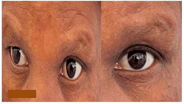
Figure 1: Before-and-after eye treatment photos showing reduced under-eye puffiness, smoother skin texture, and a brighter, refreshed appearance with improved contour and overall facial rejuvenation
| Parameter | Result at admission | Reference range |
| Haemoglobin | 11.5 g/dL | 12.0-16.0 g/dL |
| Platelets | 170 × 109/L | 150-400 × 109/L |
| Leukocytes | 3.23 × 10⁹/L | 4.00-11.00 × 10⁹/L |
| ESR | 51 mm/hour | 0-30 mm/hour |
| CRP | 2.0 mg/L | <3.0 mg/L |
| IgG | 1,093 mg/dL | 600-1,560 mg/dL |
| HIV-1 RNA (viral load) | 11,000 copies/mL | 20-10,000,000 copies/mL |
| ANA | Negative | - |
| ANCA | Negative | - |
| Hepatitis B/C | Negative | - |
| Syphilis | Negative | - |
| TB screening (IGRA) | Negative | - |
| CD4 count | 250 cells/µL | Normal >500 cells/µL |
Table 1: Blood test table of laboratory blood test
Non-pharmacological management performed included: Admission for better neurological assessment and regular neurological observations (level of consciousness, pupils, limb power), fall-risk assessment and assistance with mobilisation if weakness is significant, eye patch over one eye if binocular diplopia is distressing, physiotherapy for limb weakness and gait training, occupational therapy for activities of daily living, speech and language assessment if dysarthria or dysphagia develops, ensure adequate nutrition and hydration, patient counselling and health education.
Pharmacological management included antiretroviral therapy, antiplatelets aggregation, symptomatic and supporting therapy.
Comments and final remarks
HIV-associated vasculitis is relatively rare (reported incidence of 1-5% of advanced HIV infection) and can affect various vessel sizes and types. Some authors reported that HIV-associated vasculitis predominantly affects small and medium-calibre vessels and is more frequently seen in individuals with high viral load or very low CD4 counts. Notwithstanding, the pathogenic mechanism involves direct viral effects on the vascular endothelium, immune complex deposition, and the dysregulated immune response characteristic of HIV infection. Clinically, HIV-associated vasculitis can present with diverse manifestations, depending on the specific vessels involved and the extent of the inflammatory process [29,30]. The typical histological features are seen in Figure 2.
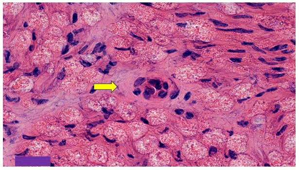
Figure 2: Shows a cross section of the blood vessel with vasculitis stained with H/E
Regarding primary HIV infection, some neurological manifestations may be seen associated with small-vessel vasculitis, direct nerve damage due to the rapid replication of the HIV virus during this phase, or reactivation of other latent viruses, such as herpes simplex or varicella zoster.
In the presented case, we thought that we were probably dealing with direct damage to the midbrain blood supply by HIV associated vasculitis.
In Figure 3, we shown the histological differences between the normal blood vessels and vasculitis. Considering the finding of midbrain damage by occlusion of small branches of the left posterior cerebral artery is most likely caused by small-vessel vasculitis induced by the autoinflammatory response associated with primary HIV infection [3].
HIV infection is characterised by progressive immune system depletion, leading to increased susceptibility to opportunistic infections and, if untreated, the development of AIDS [4]. Early detection during this phase is challenging due to the nonspecific nature of symptoms and the potential for misdiagnosis. Efforts to promote early testing and awareness are crucial to identify and manage HIV infections as soon as possible. In the context of HIV infection, there is an increased risk of immune dysregulation and exacerbation of autoimmune conditions.
In Figure 3, is shown the features of vasculitis compared with normal blood vessels.
While specific studies for autoinflammatory states in HIV infected patients are limited, it is known that HIV-related inflammation can increase the risk of autoimmune diseases such as vasculitis and other inflammatory disorders [31].
The summary of causes of vasculitis is listed in Figure 4.
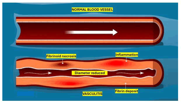
Figure 3: Shown the features of vasculitis compared with normal blood vessels
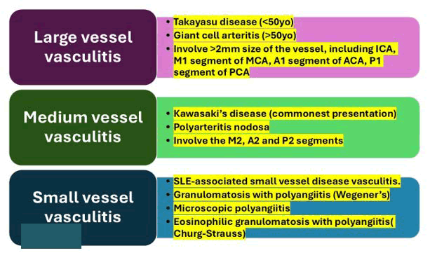
Figure 4: List the number a cause of vasculitis according with the size of the affected blood vessel
Brief comments on drug therapy
The treatment of HIV-associated vasculitis requires a dual approach: Controlling the underlying HIV infection with ART and managing the inflammatory vascular process [32].
Initiation of ART as quickly as possible is the cornerstone of drug therapy, as it helps suppress viral replication, reduce systemic inflammation, and restore immune function, thereby potentially resolving vasculitic manifestations in most people. In patients with severe or symptomatic vasculitis, adjunctive immunosuppressive therapy, such as corticosteroids, may be required to control acute inflammation and prevent end-organ damage. However, the administration of immunosuppressive drugs must be carefully monitored, as excessive immunosuppression can increase the risk of opportunistic infections, particularly in individuals with low CD4 counts, as in our reported case. In refractory cases or those involving large-vessel vasculitis, additional immunomodulatory therapies, such as methotrexate, azathioprine, or cyclophosphamide, may be considered under close monitoring [32].
Due to the risk of complications associated with both HIV infection and vasculitis, a multidisciplinary approach involving infectious disease, autoimmune disease, and vascular specialists is vital to ensure optimal drug therapy and long-term patient outcomes.
We hypothesise that the best follow-up of HIV-associated vasculitis in patients presenting with midbrain ischemic stroke relates to HIV-vasculitis, and close clinical and imaging surveillance is necessary to monitor disease progression and response to ART. For small-vessel vasculitis, such as the suspected involvement in our patient, prognosis is generally favourable with ART initiation, as immune reconstitution tends to resolve vasculitic activity over time. If the clinical picture persists or worsens, additional immunosuppressive therapy, such as corticosteroids, may be considered, though with caution due to the risk of opportunistic infections.
In patients presenting large-vessel vasculitis, particularly aortic vasculitis detected on PET-CT, long-term monitoring is crucial due to the risk of vascular complications, including vessel stenosis, aneurysm formation, or IS [33].
We also believe that for the above-cited cases, serial imaging with PET-CT or MRI angiography at regular intervals is strongly recommended to assess disease activity and detect structural changes in the great vessels. In most people, ART alone can lead to regression of vascular inflammation, but in patients with persistent vasculitis, corticosteroids or other immunosuppressants may be prescribed. The prognosis of large-vessel vasculitis in HIV remains variable, depending on the degree of vascular involvement and response to therapy. While controlled HIV infection generally leads to better outcomes, untreated or severe cases can result in lifethreatening vascular complications.
Brief comments on the role of coagulopathies in HIVIschemic stroke
It has been documented that HIV is associated with various coagulopathies such as thrombotic thrombocytopenic purpura, protein S and C deficiency, and antiphospholipid syndrome [34-36]. Decreased levels or impaired function of proteins S and C increase the risk of thrombophilia, which can lead to DVT and IS. Nonetheless, some investigations in HIV infected patients with ischemic stroke have shown that 45% of patients had protein S deficiency [35].
Other investigators reported no significant association between protein S deficiency and the occurrence of stroke in HIV positive patients, suggesting that protein S deficiency is an epiphenomenon of the HIV infection with no recognised relationship to IS [37].
Although we could not confirm it in our series, it is well proven that deficiencies in protein C and protein S were observed in IS patients with HIV; therefore, the question remains unclear whether these deficiencies are secondary events or directly caused by HIV infection. Additionally, a case-controlled study in young HIV positive IS patients demonstrated elevated levels of VWF in comparison with both uninfected and HIV-infected patients without IS [38].
Brief comments on the role of opportunistic infections
Several opportunistic infections such as neurosyphilis, tuberculosis meningitis, and varicella-zoster vasculitis may predispose people to IS. Tubercle bacillus (TB) is the most common pathogen known to cause opportunistic infections associated with HIV in our setting. In fact, IS is thought to be a complication of tuberculous meningitis [39].
HIV infection causes an immunocompromised condition that increases one’s susceptibility to secondary infections [40].
On the other hand, Varicella zoster infection remarkably elevates the risk of stroke and cerebral vasculitis in immunosuppressed patients as well [41].
In PLWH, increased meningovascular complications and neurosyphilis have been observed [42]. As mentioned before, infections are thought to trigger widespread neurovascular inflammation leading to endarteritis, a prothrombotic state, and consequent IS. The combination of arterial wall swelling and a predisposition to thrombus formation contributes to an increased risk of atherosclerosis, which leads to an IS [43].
From our comprehensive review, we could not find accurate and confident information to support the hypotheses that Candida albicans and cytomegalovirus infections are associated with HIV-patients presenting IS. Therefore, additional investigations should be done to confirm their roles in the pathogenesis of IS in HIV-positive people.
Brief comments on HIV associated vasculopathy
Despite the great advancement of ART, vascular disease remains a major cause of morbidity and mortality in the HIV-infected population and is the leading cause of IS [44].
Some investigators have reported vasculopathy in postmortem human brain samples from immunosuppressed patients [45-47].
Other authors reported that the major inflammatory damage caused by HIV infection on the arteries is localised at the level of the adventitial intima. Atherosclerosis of the large arteries and small vessels is the commonest cause of IS in HIV infected patients [48,49].
Large vessel vasculopathy with ectasia and aneurysm formation was also documented intra- and extracranially, and it was the main cause of IS and intraparenchymal haemorrhage [50,51].
Brief comments on cardioembolism and atherosclerosis
In PLWH, cardioembolic IS represents approximately 4-20% of ischemic strokes [52,53].
The main causes of cardioembolic strokes include arrhythmias, cardiac chamber abnormalities, and valve disorders. HIVpositive people have a higher risk of developing atrial fibrillation and cardiomyopathy. Additionally, bacterial and marantic endocarditis, and ischemic heart disease significantly contribute to cardioembolism [54-56].
On the other hand, HIV infection induces vascular abnormalities such as increased carotid arterial wall stiffness, carotid intimal thickness, vascular inflammation, and abnormalities in vascular compliance and distensibility in the absence of ART, which are major risk factors for atherosclerotic disease [57-59].
HIV alone can directly initiate atherogenesis by activating immune and endothelial cells, increasing the number of circulating atherogenic immune cells, and altering lipid levels and function [59]. The main elements involved in the pathogenesis of accelerated atherosclerosis in HIV infected peoples are represented in Figure 5.
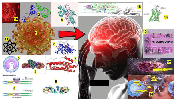
Figure 5: Graphical representation of: 1) HIV, 2) trans-activators of transcription (tat), 3) Glycoprotein-120, 4) Negative regulatory factor, 5) Moncyte chemoattractant protein, 6) Interleukin, 7) Tumor necrosis factor alpha, 8) Nuclear factor kappa-beta, 9) Monocyted, 10) CD4+T-cells, 11) Oxidative free radicals, 12) Macrophage, 13) Cell Adhesion Molecules (CAMs), 14) Chemoattract Chemokine Ligands (CCL2), 15) Thrombomodulin. Other elements non included are von Willebrand, Plasminogen activating inhibitor-1 antigen, D-Dimer
The vascular endothelium is permanently exposed to stimuli such as HIV-infected cells (monocytes, CD4+ T cells, macrophages, and circulating viruses), viral proteins released with host cell lysis and actively secreted, and virus-induced proinflammatory mediators. Therefore, these stimuli will damage the vascular endothelium, increase its permeability, and lead to extravasation of immune cells, ultimately resulting in chronic inflammation [60].
On the other hand, HIV infection trigger the synthesis of oxidative free radicals, Cell Adhesion Molecules (CAMs), and release of chemoattractant such as Chemokine Ligand 2 (CCL2) at the site of inflammation, which attracts leukocytes, and on top of that, endothelial specific coagulator molecules such as plasminogen activator inhibitor-1 antigen, tissue factor, von Willebrand factor (VWF), thrombomodulin, and d-dimer are disturbed in HIV infection, favouring a prothrombotic state, which potentially accelerates atherosclerosis [61,62].
The HIV viral proteins, the trans-activator of transcription (tat), glycoprotein-120 (gp120), and negative regulatory factor (nef) are important participants of the immune expression. The Tat and gp120 proteins increase CAM expression, induce oxidative stress, and enable leukocyte transmigration and adhesion to the endothelium [63,64].
The Tat induces the production of Monocyte Chemoattractant Protein-1 (MCP-1), which recruits monocytes to the site of infection and promotes the synthesis of Tumour Necrosis Factor Alpha (TNF-a), Nuclear Factor kappa-B (NF-?B) and Interleukin-6 (IL-6) [65,66].
gp120 boosts TNF-a synthesis and immunoglobulin secretion in B-lymphocytes and stimulates macrophage expression, along with Nef. Furthermore, Nef proteins promote the transformation of macrophages into foam cells, a process that underlies atherosclerotic transformation. Altogether, HIV viral proteins create a pro-inflammatory milieu that facilitates atherogenesis [67-69].
Brief comments on antiretroviral therapy
We hypothesised that the increased incidence of IS in HIV-positive patients is due to the increased prevalence of opportunistic infections and inflammation due to immunosuppression, and older ART regimens that predisposed patients to dyslipidaemia and lipodystrophy, as has been suggested by others [70].
However, HIV/AIDS therapy also increases the risk of IS, both directly by accelerating atherosclerosis and indirectly by increasing life expectancy [71].
Nevertheless, ART has changed the HIV treatment outcomes; low levels of viral suppression in the CNS reservoirs leading to increased risk of IS must be taken into consideration. The same authors have demonstrated that prolonged ART treatment was associated with an increased prevalence of cardiovascular disease and IS [72].
While other investigators have highlighted that immunosuppression and increased viral loads are associated with increased incidence of IS [73-75], low viral titter can induce low-grade systemic inflammation, which may further add to the risk of stroke.
Prolonged use of Protease Inhibitors (PIs) like darunavir is associated with stroke and myocardial infarction [76].
Moreover, atazanavir is associated with vascular remodelling and Nucleoside Reverse Transcriptase Inhibitor (NRTI), and abacavir is also associated with increased incidence of cardiovascular events and stroke [77].
Despite ART reducing the virulence of HIV and increasing life expectancy in PLWH, with long-term endothelial and metabolic challenges, there is a great risk of IS. Conversely, there are few studies that report ART is associated with reduced risk of IS. This is where ART functions to control the HIV infection, whereby viral suppression and improved immune function confer protection against stroke. However, prolonged exposure to ART may lead to a rise in vascular risk over time [78].
We also believe that there are challenges when dealing with the clinical manifestation of HIV-associated stroke. Improved life expectancy in treated HIV patients increases incidence traditional risk factors along with prolonged viral infection and side effects of ART. However, there are several ways to confront these challenges, starting from preventive measures to early screening of risk factors and novel therapeutic interventions.
Because of its narrow therapeutic window, stroke reperfusion therapies have led to remarkable improvement in clinical outcomes; however, a small population of patients are benefiting. ART drugs are known to be beneficial for suppressing the viral load, but their prolonged use is still not clinically proven safe for HIV patients.
Brief comments on our patient
Despite having a midbrain infarction, our patient was stable and had moderate to mild disability at hospital discharge. We treated this patient appropriately; the condition gradually improved with medical treatment and physical therapy. We reported a very rare case of IS as Weber’s syndrome related to HIV-vasculitis/atherosclerosis. This condition was caused by an ischemic lesion in the ventromedial midbrain with very severe stenosis of branches of the left posterior cerebral artery. The vascular territory of the posterior cerebral artery and its branches are shown in Figure 6.
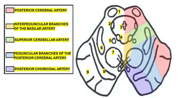
Figure 6: Shows a cross section of the midbrain with the vascular territory of different arterial branches. 1) Superior colliculus, 2) Mesecephalic nucleus of the trigeminal nerve, 3) Aqueduct of Sylvius, 4) Oculomotor nucleus, 5) Edinger Westphal nucleus, 6) Medial lemniscus, 7) Red nucleus, 8) Substantia nigra, 9) Cerebral peduncle
This syndrome is usually caused by a lack of blood supply to the brain from the paramedian mesencephalic branches (basilar), the peduncular perforating branches (posterior cerebral artery), the choroidal arteries, and the superior cerebellar artery, which supply the midbrain, resulting from an isolated occlusion of these vessels. In addition, other less common causes of this syndrome are haemorrhage, aneurysms, cavernomas, tumours, and demyelinating diseases [79].
In this syndrome, the hallmark neurological findings are crossed sensory disturbance or motor weakness and oculomotor nerve palsy [5,6]. Our reported patient had right-side motor weakness, medial gaze palsy, with anisocoria, with the left pupil (5 mm) and the right one (3 mm), due to third nerve palsy with diplopia. The patient’s condition improved gradually with medical treatment and physical therapy.
The oculomotor nuclei are located at the level of the superior colliculus in the midbrain and extend approximately 10 mm from rostral to caudal. Most of the fiber in the pupils is supplied by the Edinger Westphal nucleus in the upper midbrain, while most of the fiber in the extra-ocular eye muscles is supplied by the motor nucleus in the lower midbrain [3,4].
Pathological lesions in the lower midbrain impair extraocular muscles but not pupils, whereas lesions in the upper midbrain cause pupillary dilatation [4,5]. Most patients with Weber’s syndrome are neurologically stable [80].
Brief comments on targeting ferroptosis and necroptosis to treat stroke
We investigated the role played by different types of programmed cell death in the CNS associated with neurocysticercosis [81-84].
Now, we investigate the role played by the previously cited pathological processes in the pathophysiology of IS, which is a leading cause of death and long-term disability worldwide. It results from cerebral blood flow obstruction (ischemia) and, in some cases, the restoration of cerebral blood flow (reperfusion), triggering a cascade of pathophysiological events collectively known as the ischemic cascade and reperfusion injury, which leads to the activation of different regulated cell death types, and this review focuses on necroptosis and ferroptosis. Both pathways are closely linked to oxidative stress and contribute significantly to neuronal death and inflammation following ischemic stroke. Dysregulation of redox homeostasis, iron dyshomeostasis, glutathione depletion, and mitochondrial dysfunction are key events in neuronal damage. Understanding the interplay between oxidative stress and these pathways is crucial for developing effective neuroprotective therapies. This review highlights recent advances in understanding necroptosis and ferroptosis in ischemic stroke, proposing redox-targeted interventions as promising strategies to mitigate brain injury and improve outcomes in patients affected by this condition.
Brief comments on therapeutic drug
The rt-PA contributes to neurovascular injury during reperfusion and supports the need to better understand its interactions with regulated cell death pathways, such as ferroptosis and necroptosis. Importantly, patients who survive an IS have an increased risk of recurrent events. Prevention of a new event involves antiplatelet and anticoagulant therapy, as well as control of causal risk factors, with a focus on the underlying mechanisms [85].
As we documented before, IS represents a clinical condition characterised by the sudden onset of focal neurological deficits caused by an underlying cerebrovascular disease, being the second cause of death worldwide, only preceded by ischemic heart disease, and it is expected to remain so in 2030. Furthermore, it is the leading neurological diseasecausing disability in the world population [86,87].
To align with the main goal of acute management of IS, it is mandatory to optimise haemodynamic status and achieve early tissue reperfusion. Hemodynamic status is optimised by controlling fluid volume, blood pressure, and cardiovascular function. Tissue reperfusion is achieved with intravenous (i.v.) thrombolysis or mechanical thrombectomy [88].
Recombinant tissue Plasminogen Activator (rt-PA) is the treatment of choice for acute IS. rt-PA binds fibrin and converts plasminogen to plasmin, which degrades fibrin and dissolves the clot [89].
Alteplase is the oldest approved plasminogen activator used for ischemic stroke (1996). It is administered i.v. administered within 3 h after ischemic stroke symptoms onset with a dose of 0.9 mg/kg (maximum 90 mg) over 60 min with an initial bolus of 10% [90] and can administered in patients until 4.5 h with the same dose, but considering the following exclusion criteria: a) Older than 80 years old, b) With a National Institute of Health Stroke Scale (NIHSS) score>25, c) Who take oral anticoagulants, d) With diabetes mellitus, or e) Who have had a previous stroke [91].
Additionally, alteplase could be administered to patients who awake with stroke symptoms and have a Diffusion- Weighted Magnetic Resonance Imaging (DW-MRI) lesion smaller than 1/3 of the middle cerebral artery territory, with no change on T2-weighted Fluid-Attenuated Inversion Recovery (FLAIR) [90]. It can also be used in patients with focal neurological deficits without a differential diagnosis of stroke type. Moreover, alteplase is strictly contraindicated in patients with haemorrhagic stroke, tumours, abscesses, or any vascular malformation [91].
On the other hand, Tenecteplase at the dose of 15–25 mg according to patient weight (<60 kg to = 90 kg) in a single i.v. bolus; it’s a derivative of alteplase but with 3 amino acid substitutions, has remarkable advantages over alteplase such as a prolonged half-life, major fibrin specificity, resistance to the Plasminogen Activator Inhibitor-1 (PAI-1) and is more effective than alteplase at lysing clots in large vessels [92,93].
Another procedure to removes a clot (thrombus) obstructing the cerebral blood flow in large vessels using stent-retrieval or aspiration is known as Mechanical Thrombectomy (MT) and it has proven to be quite effective in patients within 6 h after IS symptoms onset, with a pre-stroke modified Rankin Scale (mRS) score of 0 to 1, occlusion of the internal carotid or middle cerebral artery segment 1 (M1), age = 18, NIHSS score = 6, and Alberta Stroke Program Early Computed Tomography Score (ASPECTS) = 6. Notwithstanding, MT using stent retrieval could be considered in some patients with ischemic infarct in the middle cerebral artery territory at segments 2 and 3 (M2 and M3). Moreover, MT is recommended for patients with large-vessel occlusion in the anterior circulation, with symptom onset within 6–24 h and DAWN eligibility criteria or 6–16 h and DEFUSE3 eligibility criteria [90].
The rt-PA is the main pharmacological therapy in IS, but some adverse side effects have been reported when it is administered after its therapeutic window. Some investigators have documented that rt-PA may disrupt the blood–brain barrier [94].
Trigger ischemic-to-haemorrhagic stroke transformation [95], enhance excitotoxicity [96], and amplify inflammatory [97] and proteolytic responses.
Brief comments on pathophysiology of midbrain ischemic stroke
Under physiological circumstances, cerebral blood flow (50–60 mL/100 g tissue/min) provides the oxygen and glucose necessary for normal brain function [98,99].
At the mitochondrial level in neurons, ATP is primarily produced via the citric acid cycle, providing the energy necessary to maintain the membrane potential and generate action potentials, thereby enabling neuronal communication throughout the network [100].
An action potential opens Voltage-Gated Calcium Channels (VGCCs), resulting in Ca2+ influx and promoting synaptic vesicle fusion with the plasma membrane, thereby releasing neurotransmitters such as glutamate [101].
In Figure 7, we graphically represented the elements involved in cerebral blood flow and synapses under physiological conditions.
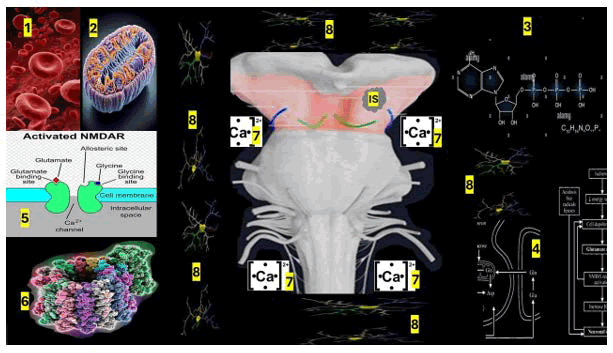
Figure 7: Schematic overview of the proposed mechanism illustrating the interaction of NMDA receptor activation, Calcium (Ca²?) signaling, mitochondrial function, neuronal networks, and molecular pathways that collectively contribute to the observed biological effects. Numbers indicate the corresponding components described in the figure
Note: 1) Reduced cerebral blood flow due to a clot/thrombus/vasculitis causing a reduction of oxygen and glucose delivery to the parenchymal tissue, promoting an ischemic core, where some biochemical alteration known as the ischemic cascade occurs. 2) Metabolism shifts from aerobic to anaerobic, leading to a decrease in ATP levels and an increase in lactate production, reducing cell pH level (Cells switch from aerobic to anaerobic respiration when oxygen supply is insufficient to meet energy demands). 3) The depletion of ATP results in an imbalance of ion gradients, due to the Na+/K+- ATPase impairment leading to increased extracellular K+ and intracellular Na+ levels, contributing to membrane depolarisation and failure of normal neurophysiological function in the affected zone. An increase in cytoplasmic Na+ reverses the Na+/Ca2+ exchanger (NCX), increasing intracellular Ca2+ levels. Additionally, membrane depolarisation opens Voltage-Gated Ca2+ Channels (VGCC), thereby increasing Ca2+ influx into the cytoplasm. ATP depletion impairs the Plasma Membrane Ca2+-ATPase (PMCA), thereby increasing cytoplasmic Ca2+ levels. 4) Depolarization of the membrane and increased intracellular Ca2+ levels lead to glutamate release (Glutamate is released from neurons via synaptic vesicles in a calcium-dependent process, acting as the primary excitatory neurotransmitter in the brain), thereby increasing its concentration in the synaptic cleft and overexpression of N-methyl- D-aspartate receptors (NMDAr). 5) Ca2+ accumulates in the postsynaptic neuron due to NMDAr activation (The NMDA receptor is a crucial ionotropic glutamate receptor involved in synaptic plasticity, learning, and memory, functioning as a coincidence detector for neuronal communication), and this is exacerbated by NCX reversal and impairment of Na+/K+-ATPase and PMCA. 6) The energy failure compromises astrocyte-mediated glutamate clearance from the synaptic space via Excitatory Amino Acid Transporters (EAAT) due to Na+ gradient alteration associated with Na+/K+-ATPase impairment. 7) The overaccumulation of Ca2+ in postsynaptic neurons promotes mitochondrial Ca2+ accumulation (Calcium ions (Ca²?) act as a universal second messenger, regulating numerous cellular processes by transiently increasing intracellular concentrations in response to stimuli), which results in mitochondrial impairment and an increase in reactive oxygen species such as superoxide anion, 8) Mitochondrial activation.
Figure 7 Graphical representation of our hypothesis on the mechanism of IS at the midbrain including the most remarkable components involved in this process. 1) Normal cerebral capillary vessels supply the necessary oxygen and glucose levels to the brain through the BBB. 2) For the production of energy, ATP, which is implicated in different cellular functions, such as maintaining the membrane potential. 3) During an action potential, the Voltage-Gated Ca2+ Channel (VGCC) opens, allowing the influx of Ca2+. This increase in Ca2+ is regulated by the Na+/Ca2+ exchanger (NCX) via the Na+ antiporter and the Plasma Membrane Ca2+-ATPase (PMCA), a mechanism that depends on ATP. In turn, Na+ levels are regulated by Na+/K+-ATPase via an ATP-dependent mechanism. 4) Increase in Ca2+ in the presynaptic neuron promotes the glutamate release, which binds to the N-methyl-D-Aspartate receptors (NMDAr), 5) Allowing the influx of Ca2+ in the postsynaptic neuron. The levels of Ca2+ are regulated by PMCA and NCX transporters as well as by mitochondrial storage. 6) Glutamate levels in the synapse are regulated by the Excitatory Amino Acid Transporters (EAAT) in a Na+-dependent mechanism in the astrocytes. 7) In a physiological condition, microglia remain resting. This information was reported by other authors [102].
A decrease in cerebral blood flow to 10–15% (4.8-8.4 mL/100 g of tissue/min) creates an area called the ischemic core, as reported by other authors. This leads to cell death within a few minutes of reduced midbrain blood flow, resulting in permanent tissue damage. Whereas reductions of 28–70% (14.1 to 35.0 mL/100 g of tissue/min) result in the penumbra zone [103].
This region of the brainstem is reversibly injured and is considered a therapeutic target to reduce permanent tissue damage. The reduction in midbrain blood flow activates the ischemic cascade, resulting in cellular injury and death, ultimately leading to brainstem tissue damage. The mechanisms involved in the ischemic cascade in the brain include cellular bioenergetic failure, excitotoxicity, oxidative stress, inflammation, and cell death [104], affecting neurons, astrocytes, oligodendrocytes, pericytes and microglia.
Here, we hypothesised that the principal mechanisms involved during midbrain ischemia include some elements that are represented in Figure 7.
Additionally, xanthine oxidase catalyses the generation of superoxide anion, which is reduced to hydrogen peroxide, which is then converted to the hydroxyl radical in an Fe2+- dependent reaction. The hydroxyl radical oxidises proteins, lipids, and nucleic acids, thereby contributing to cellular damage. Also, the accumulation of Ca2+ activates proteases, phosphatases, lipases, and endonucleases, which promote protein, lipid, and DNA damage, resulting in cell damage and necrosis, and the release of cytoplasmic contents into the extracellular space. The molecules released during necrotic cell death activate microglia (Activated microglia are the immune-responsive form of CNS-resident microglial cells, undergoing morphological and functional changes to respond to injury, infection, or pathological stimuli), which in turn release pro-inflammatory cytokines, leading to inflammation, which has been proposed by other investigators under different circumstances. Some of the elements included in this hypothesis were previously reported by other investigators under different scenarios [105].
Other investigators reported that membrane depolarisation (via increased extracellular K+ levels) activates VGCC, thereby increasing cytoplasmic Ca2+ levels [106].
Furthermore, membrane depolarisation and elevated intracellular Ca2+ levels trigger glutamate release, thereby increasing its concentration in the synaptic cleft [107].
Furthermore, energy failure compromises glutamate clearance from the synaptic space via astrocytic EAAT [108,109].
Then, increasing glutamate concentration in the synaptic space overactivated NMDAr, a-amino-3-hydroxy-5- methyl-4-isoxazolpropionic acid (AMPA), and kainite receptors. Specifically, excitotoxicity results from the overactivation of NMDAr (primarily the NR2B subunit), which induces excessive Ca2+ influx into the cytoplasm and triggers signalling pathways that ultimately lead to cell death [107].
A first peak of Ca2+ occurs immediately after capillary blood flow reduction and lasts around 26 min, followed by a second and gradual increase in Ca2+ maintained for at least 3 h. The intracellular Ca2+ level is associated with the extent of cerebral damage [110].
This overload of intracellular Ca2+ activates Ca2+-dependent enzymes, including proteases, phosphatases, lipases, endonucleases, and neuronal nitric oxide synthase, thereby leading to cellular damage and cell death [107,111].
Furthermore, mitochondria are susceptible to Ca2+ overload, which leads to dysfunction, increased ROS production, decreased mitochondrial membrane potential, release of cytochrome c into the cytoplasm, and subsequent activation of apoptosis [112-114].
Additionally, the interaction between the Transient Receptor Potential Melastatin 4 (TRPM4), an impermeable Ca2+ channel, and the NMDAr (NR2B subunit) can exacerbate excitotoxicity [115]. ROS are oxygen-derived molecules partially reduced and highly reactive, produced during normal metabolism by mitochondria, NADPH oxidase, xanthine oxidase, cytochrome P450, peroxisomes, endoplasmic reticulum, and other enzymes (± 50 different sources) [116].
Physiologically, ROS play a crucial role in cellular signalling, cell differentiation and proliferation, gene expression, epigenetic modifications, migration, angiogenesis, and regulation of vascular tone [117,118].
However, an imbalance between ROS production and antioxidant defences generates oxidative stress, in which lipids (lipoperoxidation, as indicated by Malondialdehyde (MDA) and 4-hydroxy-2-nonenal (4-HNE) levels), proteins (nitrotyrosine and carbonyl group levels), and DNA (8-hydroxy-2’-deoxyguanosine level) are oxidised. These oxidative modifications result in cellular damage and death [119,120].
ROS production is region-specific in the brain. Some investigators have documented that the brainstem and cerebellum exhibit the highest levels of ROS under resting conditions, and that these ROS are associated with glial cells. Stimulation with ATP or glutamate leads to the highest ROS production in the midbrain, whereas lipoperoxidation is highest in the hippocampus and the midbrain [121].
The continuous reduction in capillary blood flow augments ROS production for up to 100 min; however, ROS production increases at the onset of reperfusion after 60 min of ischemia [122]. Other authors reported that in cortical neuron and hippocampal cultures subjected to oxygen– glucose deprivation, ROS production happens through three mechanisms. The first rise of ROS is generated by mitochondria at 3–7 min, followed by a second increase caused by xanthine oxidase, and the third rise of ROS occurs after reoxygenation and glucose restitution (reperfusion), and mitochondria and NADPH oxidase are responsible for this last increase [123].
Superoxide radical anion increases immediately after ischemia and reaches a steady state at 180 min. In contrast, Hydrogen Peroxide (H2O2) production is maintained for up to 48 h after the ischemic insult. Restoration of cerebral blood flow (reperfusion) increases ROS production, thereby activating cell death pathways, including apoptosis, necroptosis, and ferroptosis in the penumbra zone [124].
Brief comments on primary mechanisms activated during reperfusion.
In Figure 8, most of the mechanisms of reperfusion in the midbrain after IS are represented according to our hypotheses.
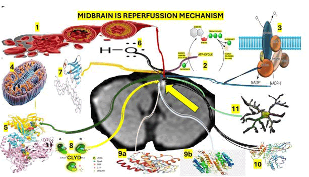
Figure 8: Shows graphically our hypothesis on the mechanism of reperfusion in the cross section at the rostral midbrain seen in MRI with IS
Note: 1) The restoration of cerebral blood flow to the penumbra zone (Restoration of cerebral flow is a critical aspect of stroke treatment, aiming to improve blood flow to the brain and potentially prevent or mitigate damage). 2) Results in a sudden oxygen and glucose supply, increasing ATP production. 3) Particularly, the increase in oxygen raises superoxide anion production by the NADPH oxidase (NADPH Oxidase (NOX) which are a family of enzymes that create Reactive Oxygen Species (ROS). These enzymes play a crucial role in various physiological processes, including immune defense, cellular signaling, and re gulation of gene expression in vascular cells. The production of ROS by NOX enzymes is tightly regulated to maintain homeostasis and prevent damage to cells. Misreg ulation of NOX can lead to various diseases, including cancer and neurodegenerative disorders. 4) Mitochondria. 5) On a smaller scale, the xanthine oxidase. Superoxide anion is reduced to other reactive oxygen species, such as hydrogen peroxide, which is then converted to the hydroxyl radical in an Fe2+-dependent reaction. Hydroxyl radical oxidises proteins and lipids (lipoperoxidation), thereby promoting cell membrane disruption and the release of cellular contents into the extracellular space, resulting in ferroptosis cell death. Hydroxyl radicals also oxidise DNA. Mitochondrial impairment promotes. 8) Cytochrome c release, resulting in intrinsic cell apoptosis. On the other hand, 10 pro-inflammatory cytokines such as Tumor Necrosis Factor alpha (TNFa) bind to its receptor, the Tumor Necrosis Factor Receptor (TNFR). 9) Leading to the recruitment of the TNFR-Associated Death Domain (TRADD), the cellular Inhibitor of Apoptosis (cIAP), the TNFR-Associated Factor (TRAF), and the Receptor-Interacting Protein 1 (RIP1) to the TNFR cytoplasmic domain, followed by the ubiquitination of RIP1 (9.a) by cIAP. Cylindromatosis (CLYD) deubiquitinates RIP1, resulting in cytoplasmic accumulation of RIP1 and TRADD, followed by their interaction with Fas-Associated via Death Domain (FADD) and Caspase 8 (9b), leading to extrinsic cell apoptosis. However, inhibition of caspase 8 leads to RIP1 autophosphorylation and subsequent RIP1-mediated phosphorylation of RIP3. 10) RIP3 phosphorylates the mixed lineage kinase domain-like protein (MLKL is a pseudokinase that acts as the key executor of necroptosis, a programmed form of cell death), promoting its oligomerization and membrane translocation, forming pore-like clusters, which result in the release of cellular content and cell death by necroptosis. 11) Microglia are activated by the cellular content release, resulting in the production of cytokines and inflammation.
In our hypothesis we also considered that occlusion/reperfusion, M1/M2 classically/alternatively activated microglia phenotypes, MLKL mixed lineage kinase domain-like protein, NAC N-acetylcysteine, NLRP3 NLR family pyrin domain-containing 3, OGD/Rx oxygen– glucose deprivation/reoxygenation, pRIP1 receptorinteracting protein kinase 1 phosphorylated, pRIP3 receptor-interacting protein kinase 3 phosphorylated, Rp reperfusion, RIP1 receptor-interacting protein kinase 1, RIP3 receptor-interacting protein kinase 3, ROS reactive oxygen species, Rx reoxygenation, SH-SY5Y human neuroblastoma cell line.
In 2013, we hypothesised the role of oxidative stress in the pathophysiology of IS, and the remarkable effect of antioxidants and their neuroprotective properties in patients presenting with IS related to racemose NCC [125,126]. M1 and M2 are early events in IS, producing proinflammatory factors. Microglial cells rise at 12 h after the ischemic insult, reaching a maximum level by 3 days [127] and remaining elevated until day 21. These immune cells could account for up to 80% of cells in the ischemic core on day 14 [128].
Nevertheless, peripheral immune cells (macrophages) are recruited to the affected brain region, followed by microglial proliferation, which begins to infiltrate the brain 24 h after the ischemic insult and peaks after 3 days. On the other hand, neutrophils increase on day 3 and remain elevated until 7 days post-injury [127].
As we cited before and has been confirmed by other authors, the damage caused by microglia and peripheral immune cells to the brain parenchyma results from their proliferation and the production and secretion of proinflammatory cytokines, which activate cell death pathways in nearby neurons [129].
On top of that, microglial cells can phagocytise damaged but viable cells, such as stressed neurons, thereby increasing neuronal loss and brain damage [130].
Brief comments on oxidative stress and cell death in midbrain stroke
Cytoplasmic Ca2+ elevation, ATP depletion, excitotoxicity, and oxidative stress lead to necrosis in the ischemic core, and this type of cell death occurs a few minutes after cerebral blood flow interruption [131,132].
Several death pathways are observed in the penumbra zone (e.g., apoptosis, necroptosis, ferroptosis, pyroptosis, autophagy, parthanatos, PANoptosis), as represented in Table 2 and supported by other investigators [133,134].
| Cell death type | Predominant zone (corepenumbra) | Approx. time course after ischemia | Mechanism |
| Necrosis | Core | 0–1 h after ischemia onset | Rapid ATP depletion → failure of ion pumps → cytotoxic edema → membrane rupture and DAMPs release |
| Apoptosis | Penumbra | 6–24 h (can extend to 24–72 h depending on model and reperfusion time) | Intrinsic (mitochondrial) and extrinsic (death receptor) caspase activation → DNA fragmentation |
| Necroptosis | Penumbra | 6–24 h, may persist up to 72 h (inflammatory context; I/R-dependent time) | RIP1–RIP3 interaction → p-MLKL and membrane permeabilization |
| Ferroptosis | Penumbra | 6–24 h, frequently linked to early reperfusion; may extend to 24–72 h | Iron-dependent lipoperoxidation due to failure of GPX4/GSH and system Xc? |
| Pyroptosis | Penumbra | 12–48 h (acute–subacute inflammatory phase) | Inflammasome activation (e.g., NLRP3) → Casp-1 → gasdermin pore formation and IL-1ß/IL-18 release |
| Autophagy-associated cell death | Penumbra | 1–24 h (early stress response; outcome depends on magnitude and duration) | Excessive or impaired autophagic flux contributes to cell demise under sustained stress |
| Parthanatos | Penumbra | 6–24 h (DNA damage and PARP-1 overactivation after oxidative/nitrosative stress) | PARP-1 overactivation → PAR accumulation → AIF nuclear translocation → large-scale DNA fragmentation |
| PANoptosis | Penumbra | 12–72 h (overlaps inflammatory and regulated death phases; model-dependent) | Assembly of PANoptosome integrating apoptotic, necroptotic, and pyroptotic machinery |
| CuproferroPANptosis | Penumbra | Hypothesis (8–48 h, frequently linked to early reperfusion; may extend to 48–72 h | Iron/cupper-dependent lipoperoxidation by unknown mechanism |
Table 2: Cell death pathways in the ischemic core and penumbra zone
We believe that the evidence identified in our review supports their relevance as potential therapeutic targets, and that both pathways are activated by oxidative stress. Also, they can increase ROS production, thereby forming a vicious cycle that exacerbates cellular damage and death, suggesting that antioxidant molecules could be beneficial tools for reducing the resulting damage [135].
Therefore, exploring their therapeutic potential might provide a promising approach for decreasing disability and mortality associated with IS.
Conclusion
Based on our comprehensive review of the medical literature, we concluded that Posterior Cerebral Artery (PCA) midbrain stroke represents a smaller proportion of IS, often results in significant neurological deficits and poses distinctive therapeutic challenges. Notwithstanding, endovascular thrombectomy confers early neurological recovery but does not improve long-term functional independence and is associated with higher risks of symptomatic haemorrhage and mortality compared with the best medical management, which has been supported by other authors recently.
As far as we know, this is the first study that graphically represent the elements involved in the pathogenesis of midbrain stroke secondary to HIV vasculitis and its mechanism of reperfusion at the rostral midbrain.
Acknowledgment
To thanks to Dr. Sibi Joshep for his collaboration on the management of our case series.
Ethics Statement
The current review does not qualify for ethical approval.
Patient Privacy
All information related to identification of patients were removed to provide complete anonymity.
Conflicts of Interest
Authors report no conflicts of interest.
References
- T.N. Nguyen, M.M. Qureshi, D. Strambo, Endovascular versus medical management of posterior cerebral artery occlusion stroke: The PLATO Study, Stroke, 54(2023):1708–1717.
[Crossref] [Google Scholar] [PubMed]
- L. Caplan, C.S. Chung, R. Wityk, New England Medical Center Posterior Circulation Stroke Registry: I. Methods, database, distribution of brain lesions, stroke mechanisms, and outcomes, J Clin Neurol, 1(2005):14–30.
[Crossref] [Google Scholar] [PubMed]
- O. Kuybu, P. Tadi, R.H. Dossani, Posterior cerebral artery stroke, StatPearls, (2023).
[Google Scholar] [PubMed]
- P.J. Martin, H.M. Chang, R. Wityk, L.R. Caplan, Midbrain infarction: Associations and aetiologies in the New England Medical Center Posterior Circulation Registry, J Neurol Neurosurg Psychiatry, 64(1998):392–395.
[Crossref] [Google Scholar] [PubMed]
- M. Sheikh Hassan, N. Osman Sidow, B.A. Adam, A.A. Adani, Superior alternating hemiplegia (Weber's syndrome): Case report, Ann Med Surg (Lond), 77(2022):103674.
[Crossref] [Google Scholar] [PubMed]
- D.I. Algin, F. Taser, S. Aydin, E. Aksakalli, Midbrain infarction presenting with Weber's syndrome and central facial palsy: A case report, Arch Neuropsychiatry, 46(2009).
- R. Abdelmasih, R. Abdelmaseih, E. Monsour, J. Reed, Isolated third nerve palsy as a rare and solo presentation of internal carotid artery dissection in a young female: A surprising finding in the angiogram, Cureus, 13(2021):e14035.
[Crossref] [Google Scholar] [PubMed]
- T. Yamaguchi, Y. Yakushiji, Bilateral paramedian thalamic infarct, Am J Med, 135(2022):e24–e25.
[Crossref] [Google Scholar] [PubMed]
- World Health Organization. HIV and AIDS, World Health Organization, (2024).
- B. Simão-Parreira, A. Costa, H. Cerveira, J. Almeida, L. Cardoso, Human immunodeficiency virus-associated vasculitis: A case report of sensorineural hearing loss, Bell's palsy, and psoriasis guttata, Cureus, 18(2026):e104705.
- A.S. Fauci, G.K. Folkers, H. Lane, Human immunodeficiency virus disease: AIDS and related disorders, Harrison's Principles of Internal Medicine, McGraw Hill, (2022).
- L.R. Ruperto, C.B. Arenzana, A.R. Marhuenda, J.I. Bernardino, Autoimmunity and HIV infection, Autoimmune disease associated with different clinical features, Elsevier, 2022:141–167.
- J.H. Schaefer, C. Stephan, C. Foerch, W. Pfeilschifter, Ischemic stroke in human immunodeficiency virus-positive patients: An increasingly age-related comorbidity, Eur Stroke J, 5(2020):252–261.
[Crossref] [Google Scholar] [PubMed]
- L.A. Benjamin, A. Bryer, H.C. Emsley, HIV infection and stroke: Current perspectives and future directions, Lancet Neurol, 11(2012):878–890.
[Crossref] [Google Scholar] [PubMed]
- L.A. Benjamin, A. Bryer, S. Lucas, Arterial ischemic stroke in HIV: Defining and classifying etiology for research studies, Neurol Neuroimmunol Neuroinflamm, 3(2016):e254.
[Crossref] [Google Scholar] [PubMed]
- K. Anders, K.D. Steinsapir, D.J. Iverson, B.J. Glasgow, L.J. Layfield, et al. Neuropathologic findings in the acquired immunodeficiency syndrome (AIDS), Clin Neuropathol, 5(1986):1–20.
[Google Scholar] [PubMed]
- M. Valdes-Sueiras, D.L. Commins, W. Yong, M. Carlson, HIV stroke risk: Evidence and implications, Ther Adv Chronic Dis, 4(2013):61–70.
[Crossref] [Google Scholar] [PubMed]
- A.I. Qureshi, R.S. Janssen, J.M. Karon, J.P. Weissman, M.S. Akbar, Human immunodeficiency virus infection and stroke in young patients, Arch Neurol, 54(1997):1150–1153.
[Crossref] [Google Scholar] [PubMed]
- W.F. Guerra, U. Tomiyasu, M.A. Verity, H.V. Vinters, The neuropathology of AIDS: UCLA experience and review, Am J Pathol, 124(1986):537–558.
[Google Scholar] [PubMed]
- A.N. Pinto, AIDS and cerebrovascular disease, Stroke, 27(1996):538–543.
[Crossref] [Google Scholar] [PubMed]
- H. Mizusawa, A. Hirano, J.F. Llena, M. Shintaku, Cerebrovascular lesions in acquired immune deficiency syndrome (AIDS), Acta Neuropathol (Berl), 76(1988):451–457.
[Crossref] [Google Scholar] [PubMed]
- J.W. Cole, A.N. Pinto, J.R. Hebel, D.W. Buchholz, C.J. Earley, Acquired immunodeficiency syndrome and the risk of stroke, Stroke, 35(2004):51–56.
[Crossref] [Google Scholar] [PubMed]
- S. Regan, S. Feske, J.B. Meigs, S.K. Grinspoon, V.A. Triant, et al. Comparison of ischemic stroke incidence in HIV-infected and non-HIV-infected patients in a US health care system, J Acquir Immune Defic Syndr, 60(2012):351–358.
[Crossref] [Google Scholar] [PubMed]
- F.C. Chow, S. Regan, M.V. Zanni, S.E. Looby, C.D. Bushnell, et al. Elevated ischemic stroke risk among women living with HIV infection, AIDS, 32(2018):59–67.
[Crossref] [Google Scholar] [PubMed]
- T.J. Henrich, S.G. Deeks, S.K. Pillai, Measuring the size of the latent human immunodeficiency virus reservoir: The present and future of evaluating eradication strategies, J Infect Dis, 215(2017):S134–S141.
[Crossref] [Google Scholar] [PubMed]
- L. Bertrand, F. Méroth, M. Tournebize, A.R. Leda, E. Sun, et al. Targeting the HIV-infected brain to improve ischemic stroke outcome, Nat Commun, 10(2019):2009.
[Crossref] [Google Scholar] [PubMed]
- S. Ismael, M.M. Khan, P. Kumar, S. Kodidela, G. Mirzahosseini, et al. HIV associated risk factors for ischemic stroke and future perspectives, Int J Mol Sci, 21(2020):5306.
[Crossref] [Google Scholar] [PubMed]
- S. Ismael, M.M. Khan, P. Kumar, S. Kodidela, G. Mirzahosseini, et al. HIV associated risk factors for ischemic stroke and future perspectives, Int J Mol Sci, 21(2020):5306.
[Crossref] [Google Scholar] [PubMed]
- L.E. Vega, L.R. Espinoza, Vasculitides in HIV infection, Curr Rheumatol Rep, 22(2020):60.
[Crossref] [Google Scholar] [PubMed]
- A. Rosenberg, P. Bailey, M. Rigamer, M. Levy, HIV-associated aortitis causing rapid development of an abdominal aortic aneurysm, Ann Vasc Surg, 66(2020):669–675.
[Crossref] [Google Scholar] [PubMed]
- C. Fujitnirun, S. Setthawatcharawanich, P. Sathirapanya, K. Phabphal, K. Limapichat, et al. Peripheral facial paralysis associated with HIV infection: A case series and literature review, Clin Neurol Neurosurg, 172(2018):124–129.
[Crossref] [Google Scholar] [PubMed]
- L.E. Vega, L.R. Espinoza, Vasculitides in HIV infection, Curr Rheumatol Rep, 22(2020):60.
[Crossref] [Google Scholar] [PubMed]
- Y. Ferfar, L. Savey, C. Comarmond, Large-vessel vasculitis in human immunodeficiency virus-infected patients, J Vasc Surg, 67(2018):1501–1511.
[Crossref] [Google Scholar] [PubMed]
- P. Vishnu, D.M. Aboulafia, Haematological manifestations of human immune deficiency virus infection, Br J Haematol, 171(2015):695–709.
[Crossref] [Google Scholar] [PubMed]
- G. Ortiz, S. Koch, J.G. Romano, A.M. Forteza, A.A. Rabinstein, Mechanisms of ischemic stroke in HIV-infected patients, Neurology, 68(2007):1257–1261.
[Crossref] [Google Scholar] [PubMed]
- C.P. Stahl, C.S. Wideman, T.J. Spira, E.C. Haff, G.J. Hixon, et al. Protein S deficiency in men with long-term human immunodeficiency virus infection, Blood, 81(1993):1801–1807.
[Google Scholar] [PubMed]
- A. Mochan, M. Modi, G. Modi, Protein S deficiency in HIV-associated ischaemic stroke: An epiphenomenon of HIV infection, J Neurol Neurosurg Psychiatry, 76(2005):1455–1456.
[Crossref] [Google Scholar] [PubMed]
- S. Allie, A. Stanley, A. Bryer, M. Meiring, M.I. Combrinck, High levels of von Willebrand factor and low levels of its cleaving protease, ADAMTS13, are associated with stroke in young HIV-infected patients, Int J Stroke, 10(2015):1294–1296.
[Crossref] [Google Scholar] [PubMed]
- D. Maher, A. Harries, H. Getahun, Tuberculosis and HIV interaction in sub-Saharan Africa: Impact on patients and programmes; implications for policies, Trop Med Int Health, 10(2005):734–742.
[Crossref] [Google Scholar] [PubMed]
- G.A. Lammie, R.H. Hewlett, J.F. Schoeman, P.R. Donald, Tuberculous cerebrovascular disease: A review, J Infect, 59(2009):156–166.
[Crossref] [Google Scholar] [PubMed]
- J. Gutierrez, G. Ortiz, HIV/AIDS patients with HIV vasculopathy and VZV vasculitis, Clin Neuroradiol, 21(2011):145.
[Crossref] [Google Scholar] [PubMed]
- L.M. Chahine, R.N. Khoriaty, W.J. Tomford, M.S. Hussain, The changing face of neurosyphilis, Int J Stroke, 6(2011):136–143.
[Crossref] [Google Scholar] [PubMed]
- F.C. Chow, V.A. Triant, Stroke in HIV, Can J Cardiol, 35(2019):280–287.
[Crossref] [Google Scholar] [PubMed]
- R.K. Heaton, D.R. Franklin, R.J. Ellis, J.A. McCutchan, S.L. Letendre, et al. HIV-associated neurocognitive disorders before and during the era of combination antiretroviral therapy: Differences in rates, nature, and predictors, J Neurovirol, 17(2011):3–16.
[Crossref] [Google Scholar] [PubMed]
- B.F. Guedes, H.R. Gomes, L.T. Lucato, P. Puglia, R. Nitrini, et al. Human immunodeficiency virus-associated vasculopathy with CNS compartmentalization of HIV-1, J Neurovirol, 21(2015):101–104.
[Crossref] [Google Scholar] [PubMed]
- J. Gutierrez, K. Menshawy, M. Gonzalez, J. Goldman, M.S.V. Elkind, et al. Brain large artery inflammation associated with HIV and large artery remodeling, AIDS, 30(2016):415–423.
[Google Scholar] [PubMed]
- W. Wang, J. Yin, B. Yuan, Z. Wang, J. Li, et al. Convulsive-like movements in posterior circulation large vessel occlusion: Associations with midbrain injury and poor outcome, CNS Neurosci Ther, 32(2026):e70980.
[Crossref] [Google Scholar] [PubMed]
- G. Ortiz, S. Koch, J.G. Romano, A.M. Forteza, A.A. Rabinstein, Mechanisms of ischemic stroke in HIV-infected patients, Neurology, 68(2007):1257–1261.
[Crossref] [Google Scholar] [PubMed]
- T.J. Allain, H. Mzinganjira, M.D. Connor, C. Smith, S. Lucas, et al. The role of human immunodeficiency virus-associated vasculopathy in the etiology of stroke, J Infect Dis, 216(2017):545–553.
[Crossref] [Google Scholar] [PubMed]
- J.A. Ake, J.C. Erickson, K.J. Lowry, Cerebral aneurysmal arteriopathy associated with HIV infection in an adult, Clin Infect Dis, 43(2006):e46–e50.
[Crossref] [Google Scholar] [PubMed]
- M. Kossorotoff, E. Touzé, S. Godon-Hardy, I. Serre, C. Mateus, et al. Cerebral vasculopathy with aneurysm formation in HIV-infected young adults, Neurology, 66(2006):1121–1122.
[Crossref] [Google Scholar] [PubMed]
- M. Hoffmann, J.R. Berger, A. Nath, M. Rayens, Cerebrovascular disease in young, HIV-infected, black Africans in the KwaZulu Natal Province of South Africa, J Neurovirol, 6(2000):229–236.
[Crossref] [Google Scholar] [PubMed]
- J. Gutierrez, K. Menshawy, M. Gonzalez, J. Goldman, M.S.V. Elkind, et al. Brain large artery inflammation associated with HIV and large artery remodeling, AIDS, 30(2016):415–423.
[Google Scholar] [PubMed]
- M. Kossorotoff, E. Touzé, S. Godon-Hardy, I. Serre, C. Mateus, et al. Cerebral vasculopathy with aneurysm formation in HIV-infected young adults, Neurology, 66(2006):1121–1122.
[Crossref] [Google Scholar] [PubMed]
- A. Pugliese, D. Isnardi, A. Saini, T. Scarabelli, R. Raddino, et al. Impact of highly active antiretroviral therapy in HIV-positive patients with cardiac involvement, J Infect, 40(2000):282–284.
[Crossref] [Google Scholar] [PubMed]
- G. Barbaro, S.D. Fisher, S.E. Lipshultz, Pathogenesis of HIV-associated cardiovascular complications, Lancet Infect Dis, 1(2001):115–124.
[Crossref] [Google Scholar] [PubMed]
- M.W. Lorenz, C. Stephan, A. Harmjanz, S. Staszewski, A. Buehler, et al. Both long-term HIV infection and highly active antiretroviral therapy are independent risk factors for early carotid atherosclerosis, Atherosclerosis, 196(2008):720–726.
[Crossref] [Google Scholar] [PubMed]
- E.C. Seaberg, L. Benning, A.R. Sharrett, J.M. Lazar, H.N. Hodis, et al. Association between human immunodeficiency virus infection and stiffness of the common carotid artery, Stroke, 41(2010):2163–2170.
[Crossref] [Google Scholar] [PubMed]
- U. Oliviero, G. Bonadies, V. Apuzzi, M. Foggia, G. Bosso, et al. Human immunodeficiency virus per se exerts atherogenic effects, Atherosclerosis, 204(2009):586–589.
[Crossref] [Google Scholar] [PubMed]
- A. Maniar, C. Ellis, D. Asmuth, R. Pollard, J. Rutledge, HIV infection and atherosclerosis: Evaluating the drivers of inflammation, Eur J Prev Cardiol, 20(2013):720–728.
[Crossref] [Google Scholar] [PubMed]
- E.A. Morgello, S. Klotman, M.E. Klotman, A. Mosoian, P.A. Lento, et al. Human immunodeficiency virus (HIV) infects human arterial smooth muscle cells in vivo and in vitro: Implications for the pathogenesis of HIV-mediated vascular disease, Am J Pathol, 172(2008):1100-1111.
[Crossref] [Google Scholar] [PubMed]
- J.F. Schved, J.C. Gris, A. Arnaud, P. Martinez, N. Sanchez, et al. von Willebrand factor antigen, tissue-type plasminogen activator antigen, and risk of death in human immunodeficiency virus 1-related clinical disease: Independent prognostic relevance of tissue-type plasminogen activator, J Lab Clin Med, 120(1992):411-419.
[Google Scholar] [PubMed]
- E.R. Kline, R.L. Sutliff, The roles of HIV-1 proteins and antiretroviral drug therapy in HIV-1-associated endothelial dysfunction, J Investig Med, 56(2008):752-769.
[Crossref] [Google Scholar] [PubMed]
- S.D. Fisher, T.L. Miller, S.E. Lipshultz, Impact of HIV and highly active antiretroviral therapy on leukocyte adhesion molecules, arterial inflammation, dyslipidemia, and atherosclerosis, Atherosclerosis, 185(2006):1-11.
[Crossref] [Google Scholar] [PubMed]
- L. Buonaguro, G. Barillari, H.K. Chang, C.A. Bohan, V. Kao, et al. Effects of the human immunodeficiency virus type 1 Tat protein on the expression of inflammatory cytokines, J Virol, 66(1992):7159-7167.
[Crossref] [Google Scholar] [PubMed]
- G. Scala, M.R. Ruocco, C. Ambrosino, M. Mallardo, V. Giordano, et al. The expression of the interleukin 6 gene is induced by the human immunodeficiency virus 1 TAT protein, J Exp Med, 179(1994):961–971.
[Crossref] [Google Scholar] [PubMed]
- P. Rieckmann, G. Poli, C.H. Fox, J.H. Kehrl, A.S. Fauci, Recombinant gp120 specifically enhances tumor necrosis factor-alpha production and Ig secretion in B lymphocytes from HIV-infected individuals but not from seronegative donors, J Immunol, 147(1991):2922-2927.
[Google Scholar] [PubMed]
- C. Lee, Q.H. Liu, B. Tomkowicz, Y. Yi, B.D. Freedman, et al. Macrophage activation through CCR5- and CXCR4-mediated gp120-elicited signaling pathways, J Leukoc Biol, 74(2003):676-682.
[Crossref] [Google Scholar] [PubMed]
- S. Swingler, A. Mann, J. Jacque, B. Brichacek, V.G. Sasseville, et al. HIV-1 Nef mediates lymphocyte chemotaxis and activation by infected macrophages, Nat Med, 5(1999):997-1003.
[Crossref] [Google Scholar] [PubMed]
- M. Bogorodskaya, F.C. Chow, V.A. Triant, Stroke in HIV, Can J Cardiol, 35(2019):280-287.
[Crossref] [Google Scholar] [PubMed]
- L.A. Benjamin, A. Bryer, H.C. Emsley, S. Khoo, T. Solomon, et al. HIV infection and stroke: Current perspectives and future directions, Lancet Neurol, 11(2012):878-890.
[Crossref] [Google Scholar] [PubMed]
- M. d'Arminio, C.A. Sabin, A.N. Phillips, P. Reiss, R. Weber, et al. Cardio- and cerebrovascular events in HIV-infected persons, AIDS, 18(2004):1811-1817.
[Crossref] [Google Scholar] [PubMed]
- H.C. Emsley, P.J. Tyrrell, Inflammation and infection in clinical stroke, J Cereb Blood Flow Metab, 22(2002):1399-1419.
[Crossref] [Google Scholar] [PubMed]
- S. Torices, T. Moreno, O.M. Osborne, S. Ramaswamy, O. Naranjo, et al. Occludin modulates HIV and ischaemic stroke response via the mitochondrial antiviral signalling pathway, Brain, 149(2026):934-950.
[Crossref] [Google Scholar] [PubMed]
- F.C. Chow, P. Bacchetti, A.S. Kim, R.W. Price, P.Y. Hsue, Effect of CD4+ cell count and viral suppression on risk of ischemic stroke in HIV infection, AIDS, 28(2014):2573-2577.
[Crossref] [Google Scholar] [PubMed]
- L. Ryom, J.D. Lundgren, W. El-Sadr, P. Reiss, O. Kirk, et al. Cardiovascular disease and use of contemporary protease inhibitors: The D:A:D international prospective multicohort study, Lancet HIV, 5(2018):e291-e300.
[Crossref] [Google Scholar] [PubMed]
- C. Bavinger, E. Bendavid, K. Niehaus, R.A. Olshen, I. Olkin, et al. Risk of cardiovascular disease from antiretroviral therapy for HIV: A systematic review, PLoS One, 8(2013).
[Crossref] [Google Scholar] [PubMed]
- C. Bavinger, E. Bendavid, K. Niehaus, R.A. Olshen, I. Olkin, et al. Risk of cardiovascular disease from antiretroviral therapy for HIV: A systematic review, PLoS One, 8(2013).
[Crossref] [Google Scholar] [PubMed]
- R.T. Pinzon, M. Afifudin, C.R.N. Ziliwu, Isolated third nerve palsy and alternate hemiparesis in brain stem stroke: A case report, Rom J Ophthalmol, 70(2026):143-146.
[Crossref] [Google Scholar] [PubMed]
- M. Rosso, S. Ramaswamy, H. Sucharew, A. Vagal, Y. Anziska, et al. Isolated third cranial nerve palsy in pituitary apoplexy: Case report and systematic review, J Stroke Cerebrovasc Dis, 30(2021):105969.
[Crossref] [Google Scholar] [PubMed]
- L.F. Ibanez Valdes, H. Foyaca Sibat, Ferropanoptosis in neurocysticercosis: A comprehensive research, Clin Schizophr Relat Psychoses, 17(2023).
- L.F. Ibanez Valdes, H. Foyaca Sibat, Novel hypotheses on the role of microglia and PANoptosis in neurocysticercosis, Clin Schizophr Relat Psychoses, 17(2023).
- L.F. Ibanez Valdes, H. Foyaca Sibat, Ferropanoptosis in neurocysticercosis: A comprehensive research, Clin Schizophr Relat Psychoses, 17(2023).
- M.N. Garcia, L.F. Ibanez Valdes, H. Foyaca-Sibat, Our hypotheses about the role of cuproferropanoptosis in neurocysticercosis and a comprehensive review, J Drug Alcohol Res, 12(2023).
- G.J. Hankey, Secondary stroke prevention, Lancet Neurol, 13(2014):178-194.
[Crossref] [Google Scholar] [PubMed]
- S.K. Powers, Ischemic stroke, Am J Med, 134(2021):1457-1468.
[Crossref] [Google Scholar] [PubMed]
- World Health Organization, Global health estimates: Life expectancy and leading causes of death and disability, Geneva (2024).
- M. Alonso de Lecinana, J.A. Egido, J. Vivancos, Guide for the treatment of acute stroke, Neurology, 29(2014):102-122.
- K.M. Bogenschutz, D.S. Fisher, G.W. Wright, Acute ischemic stroke: A guideline-based overview of evaluation and management, JAAPA, 38(2025):13-20.
[Crossref] [Google Scholar] [PubMed]
- W.J. Powers, A.A. Rabinstein, T. Ackerson, O.M. Adeoye, N.C. Bambakidis, et al. Guidelines for the early management of patients with acute ischemic stroke: 2019 update to the 2018 guidelines for the early management of acute ischemic stroke: A guideline for healthcare professionals from the American Heart Association/American Stroke Association, Stroke, 50(2019):e344-e418.
[Crossref] [Google Scholar] [PubMed]
- W. Hacke, M. Kaste, E. Bluhmki, M. Brozman, A. Davalos, et al. Thrombolysis with alteplase 3 to 4.5 hours after acute ischemic stroke, N Engl J Med, 359(2008):1317-1329.
- K.M. Bogenschutz, D.S. Fisher, G.W. Wright, Acute ischemic stroke: A guideline-based overview of evaluation and management, JAAPA, 38(2025):13-20.
[Crossref] [Google Scholar] [PubMed]
- L. Wang, M. Hao, N. Wu, S. Wu, M. Fisher, et al. Comprehensive review of tenecteplase for thrombolysis in acute ischemic stroke, J Am Heart Assoc, 13(2024):e031692.
[Crossref] [Google Scholar] [PubMed]
- M.X. Dong, Q.C. Hu, P. Shen, J.X. Pan, Y.D. Wei, et al. Recombinant tissue plasminogen activator induces neurological side effects independent on thrombolysis in mechanical animal models of focal cerebral infarction: A systematic review and meta-analysis, PLoS One, 11(2016):e0158848.
[Crossref] [Google Scholar] [PubMed]
- R.M. Dijkhuizen, M. Asahi, O. Wu, B.R. Rosen, E.H. Lo, Rapid breakdown of microvascular barriers and subsequent hemorrhagic transformation after delayed recombinant tissue plasminogen activator treatment in a rat embolic stroke model, Stroke, 33(2002):2100-2104.
[Crossref] [Google Scholar] [PubMed]
- J.P. Lopez-Atalaya, B.D. Roussel, D. Levrat, J. Parcq, O. Nicole, et al. Toward safer thrombolytic agents in stroke: Molecular requirements for NMDA receptor-mediated neurotoxicity, J Cereb Blood Flow Metab, 28(2008):1212-1221.
[Crossref] [Google Scholar] [PubMed]
- K. Shi, M. Zou, D.M. Jia, S. Shi, X. Yang, et al. tPA mobilizes immune cells that exacerbate hemorrhagic transformation in stroke, Circ Res, 128(2021):62-75.
[Crossref] [Google Scholar] [PubMed]
- K. Tsuji, T. Aoki, E. Tejima, K. Arai, S.R. Lee, et al. Tissue plasminogen activator promotes matrix metalloproteinase-9 upregulation after focal cerebral ischemia, Stroke, 36(2005):1954-1959.
[Crossref] [Google Scholar] [PubMed]
- R. Leigh, L. Knutsson, J. Zhou, P.C.M. van Zijl, Imaging the physiological evolution of the ischemic penumbra in acute ischemic stroke, J Cereb Blood Flow Metab, 38(2018):1500-1516.
[Crossref] [Google Scholar] [PubMed]
- M. Belanger, I. Allaman, P.J. Magistretti, Brain energy metabolism: Focus on astrocyte-neuron metabolic cooperation, Cell Metab, 14(2011):724-738.
[Crossref] [Google Scholar] [PubMed]
- J.S. Dittman, Taking a closer look at the synapse, Proc Natl Acad Sci U S A, 121(2024):e2412457121.
[Crossref] [Google Scholar] [PubMed]
- S.M. Bautista-Perez, C.A. Silva-Islas, R. Sanchez-Thomas, A. Figueroa, D. Barrera-Oviedo, et al. Targeting ferroptosis and necroptosis to treat stroke, Neurochem Res, 51(2026):135.
- E. Bandera, M. Botteri, C. Minelli, A. Sutton, K.R. Abrams, et al. Cerebral blood flow threshold of ischemic penumbra and infarct core in acute ischemic stroke, Stroke, 37(2006):1334-1339.
[Crossref] [Google Scholar] [PubMed]
- R. Brouns, P.P. De Deyn, The complexity of neurobiological processes in acute ischemic stroke, Clin Neurol Neurosurg, 111(2009):483-495.
[Crossref] [Google Scholar] [PubMed]
- S.M. Bautista-Perez, C.A. Silva-Islas, R. Sanchez-Thomas, A. Figueroa, D. Barrera-Oviedo, et al. Targeting ferroptosis and necroptosis to treat stroke, Neurochem Res, 51(2026):135.
- J.I. Luoma, B.G. Kelley, P.G. Mermelstein, Progesterone inhibition of voltage-gated calcium channels is a potential neuroprotective mechanism against excitotoxicity, Steroids, 76(2011):845-855.
[Crossref] [Google Scholar] [PubMed]
- Z. Shen, M. Xiang, C. Chen, F. Ding, Y. Wang, et al. Glutamate excitotoxicity: Potential therapeutic target for ischemic stroke, Biomed Pharmacother, 151(2022):113125.
[Crossref] [Google Scholar] [PubMed]
- J.I. Wadiche, S.G. Amara, M.P. Kavanaugh, Ion fluxes associated with excitatory amino acid transport, Neuron, 15(1995):721–728.
[Crossref] [Google Scholar] [PubMed]
- D.B. Kirdajova, J. Kriska, J. Tureckova, M. Anderova, Ischemia-triggered glutamate excitotoxicity from the perspective of glial cells, Front Cell Neurosci, 14(2020):51.
[Crossref] [Google Scholar] [PubMed]
- A.N. Nelson, M.S. Calhoun, A.M. Thomas, J.L. Tavares, D.M. Ferretti, et al. Temporal progression of excitotoxic calcium following distal middle cerebral artery occlusion in freely moving mice, Front Cell Neurosci, 14(2020):566789.
[Crossref] [Google Scholar] [PubMed]
- K. Szydlowska, M. Tymianski, Calcium, ischemia and excitotoxicity, Cell Calcium, 47(2010):122-129.
[Crossref] [Google Scholar] [PubMed]
- L. Schild, G. Keilhoff, W. Agustin, G. Reiser, F. Striggow, Distinct Ca²? thresholds determine cytochrome C release or permeability transition pore opening in brain mitochondria, FASEB J, 15(2001):565-567.
[Crossref] [Google Scholar] [PubMed]
- J. Li, X. Ma, W. Yu, Z. Lou, D. Mu, et al. Reperfusion promotes mitochondrial dysfunction following focal cerebral ischemia in rats, PLoS One, 7(2012):e46498.
[Crossref] [Google Scholar] [PubMed]
- S.S. Andrabi, M. Ali, H. Tabassum, S. Parveen, S. Parvez, Pramipexole prevents ischemic cell death via mitochondrial pathways in ischemic stroke, Dis Model Mech, 12(2019):dmm033860.
[Crossref] [Google Scholar] [PubMed]
- J. Yan, C.P. Bengtson, B. Buchthal, A.M. Hagenston, H. Bading, Coupling of NMDA receptors and TRPM4 guides discovery of unconventional neuroprotectants, Science, 370(2020):eaay3302.
[Crossref] [Google Scholar] [PubMed]
- L.F. Ibanez Valdes, H. Foyaca Sibat, The oxidative stress in neurocysticercosis, J Drug Alcohol Res, 13(2024).
- H. Sies, D.P. Jones, Reactive oxygen species (ROS) as pleiotropic physiological signalling agents, Nat Rev Mol Cell Biol, 21(2020):363-383.
[Crossref] [Google Scholar] [PubMed]
- C. Lennicke, H.M. Cocheme, Redox metabolism: ROS as specific molecular regulators of cell signaling and function, Mol Cell, 81(2021):3691-3707.
[Crossref] [Google Scholar] [PubMed]
- P.D. Ray, B.W. Huang, Y. Tsuji, Reactive oxygen species (ROS) homeostasis and redox regulation in cellular signaling, Cell Signal, 24(2012):981-990.
[Crossref] [Google Scholar] [PubMed]
- L.F. Ibanez Valdes, H. Foyaca Sibat, The role of oxidative stress in neurocysticercosis: A comprehensive research, Clin Schizophr Relat Psychoses, 17(2023).
- A.Y. Vinokurov, O.A. Stelmashuk, P.A. Ukolova, E.A. Zherebtsov, A.Y. Abramov, Brain region specificity in reactive oxygen species production and maintenance of redox balance, Free Radic Biol Med, 174(2021):195-201.
[Crossref] [Google Scholar] [PubMed]
- O. Peters, T. Back, U. Lindauer, C. Busch, D. Megow, et al. Increased formation of reactive oxygen species after permanent and reversible middle cerebral artery occlusion in the rat, J Cereb Blood Flow Metab, 18(1998):196-205.
[Crossref] [Google Scholar] [PubMed]
- A.Y. Abramov, A. Scorziello, M.R. Duchen, Three distinct mechanisms generate oxygen free radicals in neurons and contribute to cell death during anoxia and reoxygenation, J Neurosci, 27(2007):1129-1138.
[Crossref] [Google Scholar] [PubMed]
- B. Zhai, W. Hu, R. Hao, W. Ni, Z. Liu, Development of a ratiometric two-photon fluorescent probe for imaging of hydrogen peroxide in ischemic brain injury, Analyst, 144(2019):5965-5970.
[Crossref] [Google Scholar] [PubMed]
- H.F. Sibat, L.F.I. Valdés, Uncommon clinical manifestations of cysticercosis, in: H.F. Sibat (Ed.), Novel Aspects on Cysticercosis and Neurocysticercosis, InTech, (2013).
- L.F.I. Valdés, H.F. Sibat, The role of oxidative stress in neurocysticercosis: A comprehensive research, Clin Schizophr Relat Psychoses, 17(2023).
- M. Gelderblom, F. Leypoldt, K. Steinbach, D. Behrens, C.U. Choe, et al. Temporal and spatial dynamics of cerebral immune cell accumulation in stroke, Stroke, 40(2009):1849-1857.
[Crossref] [Google Scholar] [PubMed]
- M.Q. Jiang, Y.Y. Zhao, W. Cao, Z.Z. Wei, X. Gu, et al. Long-term survival and regeneration of neuronal and vasculature cells inside the core region after ischemic stroke in adult mice, Brain Pathol, 27(2017):480-498.
[Crossref] [Google Scholar] [PubMed]
- X. Hu, P. Li, Y. Guo, H. Wang, R.K. Leak, et al. Microglia/macrophage polarization dynamics reveal novel mechanism of injury expansion after focal cerebral ischemia, Stroke, 43(2012):3063-3070.
[Crossref] [Google Scholar] [PubMed]
- W. Chen, Y. Zhang, X. Zhai, L. Xie, Y. Guo, et al. Microglial phagocytosis and regulatory mechanisms after stroke, J Cereb Blood Flow Metab, 42(2022):1579-1596.
[Crossref] [Google Scholar] [PubMed]
- A. Rami, D. Kögel, Apoptosis meets autophagy-like cell death in the ischemic penumbra: Two sides of the same coin?, Autophagy, 4(2008):422-426.
[Crossref] [Google Scholar] [PubMed]
- J. Puyal, A. Vaslin, V. Mottier, P.G.H. Clarke, Postischemic treatment of neonatal cerebral ischemia should target autophagy, Ann Neurol, 66(2009):378-389.
[Crossref] [Google Scholar] [PubMed]
- A. Degterev, Z. Huang, M. Boyce, Y. Li, P. Jagtap, et al. Chemical inhibitor of nonapoptotic cell death with therapeutic potential for ischemic brain injury, Nat Chem Biol, 1(2005):112-119.
- Y. Cui, Y. Zhang, X. Zhao, L. Shao, G. Liu, et al. ACSL4 exacerbates ischemic stroke by promoting ferroptosis-induced brain injury and neuroinflammation, Brain Behav Immun, 93(2021):312-321.
[Crossref] [Google Scholar] [PubMed]
- U. Alam, R. Ahmed, S. Rath, F. Khattak, M.A. Asif, et al. Endovascular therapy versus best medical treatment in posterior cerebral artery stroke: A systematic review and meta-analysis, Brain Behav, 16(2026):e71194.
[Crossref] [Google Scholar] [PubMed]
Copyright: © 2026 Lourdes de Fatima Ibanez Valdes, et al. This is an open access article distributed under the terms of the Creative Commons Attribution License, which permits unrestricted use, distribution and reproduction in any medium, provided the original work is properly cited.