
Research Article: Journal of Drug and Alcohol Research (2026) Volume 15, Issue 6
Neuropsychiatric Manifestations due to Lack of Vitamin B5, B7, C, D, Iron, Magnesium, Zinc and Homocysteine. A Drug Management and Comprehensive Review
Sandisiwe Kema1, Lourdes de Fatima Ibanez Valdes2, Sibi Joseph2 and Humberto Foyaca Sibat2*2Department of Internal Medicine and Therapeutics, Nelson Mandela Academic Hospital, Walter Sisulu University, South Africa
Humberto Foyaca Sibat, Department of Internal Medicine and Therapeutics, Nelson Mandela Academic Hospital, Walter Sisulu University, South Africa, Email: humbertofoyacasibat@gmail.com
Received: 13-Apr-2026, Manuscript No. JDAR-26-192024; Editor assigned: 15-Apr-2026, Pre QC No. JDAR-26-192024 (PQ); Reviewed: 29-Apr-2026, QC No. JDAR-26-192024; Revised: 17-Jun-2026, Manuscript No. JDAR-26-192024 (R); Published: 24-Jun-2026, DOI: 10.4303/JDAR/236517
Abstract
Introduction: The role of certain micronutrients, including zinc, iron, vitamins A, D, C and B12, among others, in synergistically and individually ensuring the proper functioning of our complex immune network has become increasingly evident. Recently, some authors have established that ageing and insufficient plasma levels of water-soluble vitamins are both associated with immune decline and chronic inflammation. Therefore, a proper diet and supplementation with vitamins B1 –B12 and vitamin C will counteract these ageing-related immune alterations by supporting immune cell metabolism and reducing susceptibility to infections.
Methods: This review examines the neuropsychiatric manifestations of nutritional deficiencies. PubMed/Medline and Cochrane review databases were searched using the term and Boolean operators: (“nutritional deficiency” OR vitamin deficiency OR mineral deficiency) AND (“neuropsychiatric” OR psychiatric OR cognitive OR depression OR psychosis OR dementia OR anxiety). The search covered publications from January 2020 to June 2026. Studies were eligible for inclusion if they were peer-reviewed articles, systematic reviews, narrative reviews, or original research articles. Articles not available in English, duplicate publications, and studies not primarily focused on FND were excluded. The systematic review performed in this study followed the guidelines recommended by PRISMA (2020 statement).
Results: A total of 924 records were identified through the database search with the search terms. 214 Duplicates were removed. Remaining records underwent title and abstract screening, and 397 were excluded. Articles were excluded if they were not focused on neuropsychiatry, not nutritional deficiency-related or did not meet study eligibility. 149 full-text articles were sought for full-text retrieval. 52 records were further excluded because the full text could not be accessed, and 18 were excluded because they could not be fully translated into English. Ultimately, 94 studies were included for this review.
Conclusion: Nutritional deficiencies may manifest as neuropsychiatric symptoms. Pathophysiology is multifactorial, including impairment to cerebral energy metabolism, disruption of monoamine neurotransmitter synthesis, glutamatergic excitotoxicity through NMDA receptor dysregulation, oxidative stress and loss of antioxidant defence, neuroinflammation, and compromised myelination. Neuropsychiatric symptoms may precede the somatic and systemic manifestations associated with nutritional deficiency. In many cases, these neuropsychiatric features are incorrectly attributed to primary psychiatric disorders. Severe and life-threatening neuropsychiatric syndromes may be missed, leading to increased morbidity and mortality.
Keywords
Introduction
The role of certain micronutrients, including zinc, iron, vitamins A, D, C and B12, among others, in synergistically and individually ensuring the proper functioning of our complex immune network has become increasingly evident. Recently, some authors have established that ageing and insufficient plasma levels of water-soluble vitamins are both associated with immune decline and chronic inflammation. Therefore, a proper diet and supplementation with vitamins B1-B12 and vitamin C will counteract these ageing-related immune alterations by supporting immune cell metabolism and reducing susceptibility to infections. Despite the established relevance of micronutrients in the human diet, specific immunomodulatory properties of water-soluble vitamins in the geriatric population remain insufficiently characterised. Due to high metabolic turnover, interindividual differences in dietary intake and malabsorption, and limited storage capacity, the elderly represents a vulnerable cohort for subclinical deficiencies that might contribute to the progression of immunosenescence [1].
Vitamin B5 plays an important role in CoA synthesis and contributes to immune responses by elevating levels of acute-phase proteins [2].
The broad immunomodulatory spectrum of B5 includes enhancement of CD8+ cytotoxic T cell differentiation and anticancer immunosurveillance, as well as Th1- and Th17 cell differentiation and macrophage maturation, among others [3].
General immunologic effect of vitamin B7-consumption includes anti-inflammatory response as well as the regulation of immune-system related processes involving T-cell cytotoxicity or susceptibility to infections [4,5].
Kuroishi et al., showed that reduced biotin levels exacerbate the allergic response to nickel in mice, accompanied by increased IL-1β production, suggesting that vitamin intake might be of interest in the context of inflammationassociated metal allergies in humans as well [6].
Moreover, vitamin B7 binding to biotinylated histones reduces NF-κB gene expression. In turn, low levels of vitamin B7 correspond to a reduced activation of AMPK in human monocyte-derived dendritic cells, which enhances pro-inflammatory responses, e.g., the release of cytokines such as TNF-α, IL-12p40, IL-23, and IL-1β [7].
Unfortunately, there are few in vivo studies on the immunestimulating effects of vitamin B7. Lower levels of biotin result in human monocyte-derived dendritic cell activation and, therefore, pro-inflammatory reactions, as well as Th1- and Th17-induced pro-inflammatory responses [8].
Biotinidase Deficiency (BD) is an autosomal recessive disorder that disrupts biotin recycling and multiple carboxylase-dependent pathways. BD deficiency exhibits unique metabolic and lipidomic patterns reflecting longterm compensatory mechanisms, underscoring the value of combined omics approaches for understanding diseasespecific homeostasis and informing personalised follow-up strategies [9].
Neurodegenerative disorders, including Alzheimer’s disease, Parkinson’s disease, multiple sclerosis, and amyotrophic lateral sclerosis, represent the biggest global health burden and share convergent pathogenic mechanisms, such as mitochondrial dysfunction, oxidative stress, neuroinflammation, calcium imbalance, and neuronal loss. Nevertheless, ALA and biotin modulate key cellular pathways implicated in neurodegeneration, including mitochondrial metabolism, redox homeostasis, inflammatory signalling, and neurovascular function and provide beneficial effects on mitochondrial efficiency, oxidative stress, and neuroinflammatory markers. ALA and biotin exhibit mechanistic convergence across pathways relevant to neurodegeneration and generally favourable safety profiles [10]. Sabuti et al., recently reported that B7 deficiency alters the expression profile of colonic microRNAs, possibly contributing to alterations in the expression of proteins involved in the maintenance of colonic physiology and inflammation [11]. Chronic exposure to Manganese (Mn) induces manganism and has been widely implicated as a contributing environmental factor to Parkinson’s Disease (PD), while metabolomics analysis of the brain and body tissues of some flies at an early stage of toxicity identified systemic changes in the metabolism of biotin (also known as vitamin B7) in Mntreated groups. Biotin supplementation alleviates the pathological phenotypes of 3 standard fly models of PD and protects against Mn-induced mitochondrial dysregulation, cytotoxicity, and neuronal loss. Finally, analysis of gene expression for biotin-related proteins in PD patients revealed increased expression of biotin transporters in the substantia Nigra compared to healthy controls, suggesting a potential role for altered biotin metabolism in PD. Together, our findings identified changes in biotin metabolism as underlying Mn neurotoxicity and Parkinsonian pathology in flies, for which dietary biotin supplementation was preventative [12]. Vitamin C, also known as ascorbic acid, is a vital micronutrient and plays an essential role as a cofactor for enzymes, and is involved in collagen synthesis, neurotransmitter production, and enhancing non-heme iron absorption [13]. Vitamin C also acts as a strong antioxidant, protecting the body against oxidative stress by neutralising free radicals, other ROS, and DNA mutations induced by oxidative stress [14]. Different types of immune cells contain high intracellular levels of vitamin C, which exhibit protective properties. While it shows immunomodulatory and immunostimulant effects, a deficiency is clearly associated with higher susceptibility to infections. The effects of vitamin C are broad, including improvements in the innate and adaptive immune responses and increased lymphocyte proliferation, including NK cell activity [1].
There is remarkable worldwide variation in recommended vitamin C (Ascorbic acid) intake levels. In America, the recommendation is 90 mg/d for men and 75 mg/d for women, while in the UK, the current recommendation – established in 1991 – is only 40 mg/d for adults. Nonetheless, such results challenge the assumption that 40 mg/d is universally adequate to maintain full health. We also highlight that the UK recommendations were narrowly focused on preventing dermatological symptoms of scurvy, despite strong evidence – even at the time that vitamin C deficiency can also cause cardiac dysfunction and greater morbidity due to respiratory infections. Hemilä and Chalker conclude that the current UK vitamin C recommendation should be re-evaluated considering controlled trial evidence and broader clinical outcomes [15].
Winkler and collaborators reported that vitamin C deficiency can mimic hematologic and dermatologic conditions and highlight the importance of considering vitamin C deficiency as a potential aetiology of bleeding, even in high-income countries [16].
Ascorbate significantly contributes to a properly maintained immune status in the elderly, through strong antioxidative activities, including counteracting oxidative damage by actively accumulating the vitamin in leukocytes and promoting anti-inflammatory responses in cell-mediated immunity, thereby directly impacting the inflammaging phenotype [1].
For a long time, vitamin D has been a cornerstone of preventive medicine. However, recent investigations did not replicate the broad benefits previously observed in epidemiological studies, mainly regarding fracture and fall prevention in vitamin D-replete populations [17]. On the other hand, vitamin D deficiency is a common global health problem and remains highly prevalent in Türkiye, where limited food fortification and heterogeneous clinical practices contribute to variability in testing and supplementation strategies. Yavuz and collaborators reported that higher individualised doses of vitamin D should be considered for people with obesity, malabsorption, and certain medical conditions that impair vitamin D metabolism [18].
Iron deficiency is the most widespread nutritional deficiency worldwide, and it is the primary cause of anaemia, particularly in low- and middle-income countries and it has a multifactorial aetiology including complex interactions between genetic factors such as the Transmembrane Protease Serine 6 (TMPRSS6) rs855791 variant, which encodes matriptase-2, a protein involved in regulating hepcidin expression, and non-genetic factors, including sociodemographic, iron intake, menstrual patterns and nutritional status. Fenty et al., reported that the interaction between the TMPRSS6 rs855791 variant and nongenetic factors contributes to the risk of iron deficiency among female medical students in Yogyakarta, Indonesia [19].
Zinc is a micronutrient crucial for taste perception and can be administered to patients with dysgeusia undergoing oncology therapy. However, overconsumption of zinc can lead to copper deficiency, which is likely underrecognised and can present as fatigue, nausea, anaemia, and myelopathy [20].
Al-Musharaf et al., assessed the associations between serum and dietary Mg, Zn, and Cu levels and sleep quality in Saudi adults and found that in Saudi people, serum and dietary Mg levels were associated with poor sleep, particularly in males, while the serum Zn concentration exhibited a modest inverse association at higher levels [21].
It’s well known that zinc modulates protein expression and cytokine production, while ZIP8 facilitates IFN-γ production by increasing the intracellular zinc levels. Olah et al., reported that elderly people have a lower zinc status and IFN-γ levels. Zinc-deficient elderly participants received zinc aspartate supplementation for approximately 7 days, resulting in increased serum zinc levels, IFN-γ production, and a trend toward increased ZIP8 expression. Therefore, zinc supplementation in the elderly links zinc status to IFN-γ production, particularly through ZIP8 expression levels [22].
Nutritional deficiencies are common after Roux-en-Y Gastric Bypass (RYGB) surgery, with a severe zinc deficiency being a diagnostic challenge, given the non-specific skin findings and associated metabolic derangements. In some cases, presenting skin rash after RYGB, zinc deficiency is an important differential diagnosis [23].
Dietary zinc deficiency is a major risk factor for infection, including pneumonia, worldwide, and hospitalised patients at risk for A. baumannii infection have increased rates of zinc deficiency [24]. Type 2 Diabetes Mellitus (T2DM) is remarkably related to poor dietary habits and nutritional imbalances. Nayyar and colleagues investigated the role of zinc and chromium in T2DM pathogenesis through a combination of bioinformatics analysis, molecular docking, trace element profiling, and gene expression validation and reported markedly lower serum concentration of zinc and chromium in T2DM patients as compared to controls being the level of zinc significantly lower in T2DM patients with nephropathy and chromium level was significantly were lower in T2DM patients with stroke [25].
Hypomagnesaemia is associated with ventricular arrhythmias and may be a risk factor for heart failure, coronary artery disease and atrial fibrillation in the general population, while pregnancy is associated with a progressive physiological fall in serum magnesium concentration. In 2025, Morton reported three cases presenting dilated cardiomyopathy in the peripartum period with hypomagnesaemia and considered that Mg deficiency may be a factor in the development of dilated cardiomyopathy in pregnancy [26].
Magnesium restriction accelerates gut ageing in old but not in young mice and aggravates colitis severity, and Mg administration can protect people against Crohn’s disease, ulcerative colitis, irritable bowel syndrome, and diverticular disease. These findings identify Mg homeostasis as a key regulator of gut health and highlight Mg supplementation as a potential strategy to counteract age-related gut dysfunction [27].
Disturbances in one-carbon metabolism and Homocysteine (Hcy) regulation are involved in Alzheimer’s Disease (AD) and Parkinson’s Disease (PD), yet direct evidence from human brain tissue and the contribution of genetic variation remain limited. We investigated whether B-vitamin-related metabolic deficits and polymorphisms in one-carbon metabolism pathways contribute to cognitive impairment in AD and PD. Nonetheless, dementia in AD and PD is characterised by convergent B-vitamin deficiencies and genetic susceptibilities that disrupt Hcy metabolism. The same authors argue that it’s a mechanistic explanation for levodopa-related Hcy accumulation in vulnerable PD subjects and identify B-vitamin supplementation as a potentially modifiable factor relevant to cognitive decline [28].
Schizophrenia (SCZ) is a serious mental illness linked to neurobiological problems, such as Hyperhomocysteinaemia (HHC), increased Butyrylcholinesterase (BChE) activity, and a pro-oxidant status. Naifar and colleagues, made a case-control study finding a valuable insights into the intricate cholinergic, oxidative, and inflammatory processes linked to SCZ, which further exacerbate the clinical symptoms into a vicious cycle and concluded that increased Hcys levels happens mainly as a consequence of folate and vitamin B12 deficiencies rather than a direct pathogenic factor, underlining the need for integrated biomarker-based and nutritional approaches to improve patient outcomes [29].
Materials and Methods
This review examines the neuropsychiatric manifestations of nutritional deficiencies. PubMed/Medline and Cochrane review databases were searched using the term and Boolean operators: (“nutritional deficiency” OR vitamin deficiency OR mineral deficiency) AND (“neuropsychiatric” OR psychiatric OR cognitive OR depression OR psychosis OR dementia OR anxiety).
The search covered publications from January 2019 to June 2026. Studies were eligible for inclusion if they were peerreviewed articles, systematic reviews, narrative reviews, or original research articles. Articles not available in English, duplicate publications, and studies not primarily focused on the topic were excluded.
To provide an accurate assessment of this search, the corresponding author used a QUADAS-2 evaluation to determine that the risk of bias was low/moderate for almost all publications, and we considered the substantial technical differences observed across diagnostic protocols used in several studies. Notably, in some publications, small and mixed cohorts were analysed, including different types of diagnostic procedures, resulting in fewer cases examined under the same protocol.
Search strategy
The search covered publications from January 2019 to June 2026. Studies were eligible for inclusion if they were peerreviewed articles, systematic reviews, narrative reviews, or original research articles. Articles not available in English, duplicate publications, and studies not primarily focused on the topic were excluded.
We searched the medical literature following PRISMA guidelines. We used these Boolean terms: “Central neuropathic pain” AND “pathogenesis of neuropathic pain” OR immunopathogenesis OR diagnosis OR outcomes) AND (systematic review OR clinical study OR cohort review). We systematically searched the mentioned databases to identify articles on the cited issues. Only English-language articles were selected. Editorials, letters to the editor, preclinical studies, and conference proceedings were excluded.
Selection of study
The first author screened abstracts and titles, while others independently assessed full texts for eligibility. Publications lacking a clear diagnostic protocol, analysis, complete data, or specifics on patient numbers or AE treatment were excluded.
Selection criteria
Inclusion criteria: Articles with detailed pathogenesis, clinical features, and CNP demographic data.
Exclusion criteria were:
• Inaccessible full text.
• Articles not addressing pathogenesis or neurotransmitter for NP.
• Lack of relevant clinicopathological data.
• Non-original studies (editorials, letters, conference proceedings, book chapters).
• Non-English publications.
Data extraction and quality assessment
Study quality was rated as good, poor, fair, or reasonable according to NIH and QUADAS-2 criteria. All authors conducted separate quality evaluations, resolving disagreements through discussion and consensus. Data collection, extraction and bias assessment All abstracts and titles meeting the inclusion criteria were reviewed by the first and other authors to collect relevant information for the review. For each selected publication, data on gender, age, publication year, country, study type, total cases, and NP patient treatment were collected. Data from eligible publications were entered into an updated Excel spreadsheet.
Outcome measures
We planned to select the most relevant publications on the NPM of vitamin B complex deficiencies, their pathophysiology, and their therapy. This investigation also sought to identify novel theories on the role of neurotransmitters in neural synaptic transmission and the modulation of central NP.
Statistical analysis
Statistical analysis was performed using XLSTAT (add-on for Microsoft Excel, version 2021.4.1, Addinsoft SARL and RStudio (version 4.3.1, https://www.rstudio.com/).
Results and Discussion
Literature search
A total of 924 records were identified through the database search with the search terms. 214 duplicates were removed. Remaining records underwent title and abstract screening, and 397 were excluded. Articles were excluded if they were not focused on neuropsychiatry, not nutritional deficiencyrelated or did not meet study eligibility. 149 full-text articles were sought for full-text retrieval. 52 records were further excluded because the full text could not be accessed, and 18 were excluded because they could not be fully translated into English. Ultimately, 94 studies were included for this review (Figure 1).
Comments
The chemical structures for the vitamin discussed below are graphically represented in Figure 2.
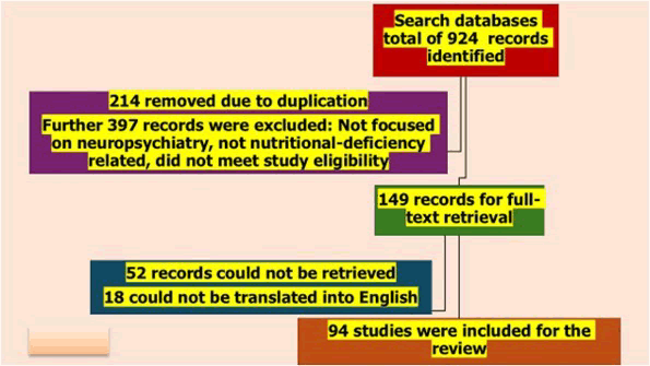
Figure 1: Flow diagram of functional neurological disorder review
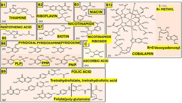
Figure 2: Illustrates chemical structures of the water-soluble vitamins B1, B2, B3, B5, B6, B7, B9, B12, C and its immunological properties, will be discussed thoroughly in the following paragraph
Brief comments on vitamin B5 (Pantothenic acid)
Vitamin B5 (Pantothenic acid) is a water-soluble B-complex vitamin [30,31]. It is an important precursor for Coenzyme A (CoA) and Acyl Carrier Protein (ACP) [30,31]. It is important for cellular energy metabolism and multiple neurochemical pathways [30].
Vitamin B5, also known as pantothenic acid (from the Greek “pantos,” meaning “everywhere”), is widely distributed in both plant and animal foods [30,31]. Pantothenic acid deficiency is rare in humans; emerging evidence links cerebral pantothenic acid depletion and impaired CoA metabolism to neurodegenerative and neuropsychiatric conditions.
Sources: Foods rich in pantothenic acid include meat, organ meats, eggs, seafood, cheese, mushrooms, legumes, whole grains, vegetables, broccoli, chickpeas, avocados, wholegrain cereals, nuts, sunflower seeds, and yeast [30,31].
Food processing, refining grains, canning, freezing, can significantly reduce pantothenic acid content [30]. Vitamin B5 can also be obtained from intestinal bacteria; the extent of this contribution is unknown [30,32]. Because deficiency is uncommon, formal recommended dietary allowances have not been established [30]. The proposed adequate intake values are 5 mg/day for adolescents and adults, 6 mg/day in pregnancy, and 7 mg/day in lactation [30].
Pharmacokinetics and pharmacodynamics: Pantothenic acid is absorbed in the small intestine primarily via a saturable sodium-dependent multivitamin transporter at physiological concentrations [30,31]. The uptake occurs by passive diffusion. After absorption, it circulates largely unbound and is taken up by tissues where it is converted to CoA through a multi-step pathway [30,31]. Pantothenic Acid Kinase 2 (PANK2) catalyses the initial step of phosphorylation of pantothenic acid to 4’-phosphopantothenic acid. The largest pools of pantothenic acid are in the form of CoA in mitochondria [30].
CoA and ACP function as acyl-group carriers in hundreds of metabolic reactions, including acetyl-CoA formation for the citric acid cycle, lipid synthesis, and acetylation reactions that regulate gene expression and protein function [30]. Excess pantothenic acid and its metabolites are excreted renally. Like other water-soluble vitamins, body stores are limited, necessitating continuous dietary supply [30,31].
Functions and pathophysiology: Forms an important part of Coenzyme A (CoA) and Acyl Carrier Protein (ACP) [30]. About 4% of cellular enzymes use CoA or its derivatives [33]. ACP is needed in fatty acid synthesis [33].
Acetyl-CoA plays many vital roles in brain metabolism. It is essential for mitochondrial energy production, various synthetic processes, and cholinergic neurons in the brain. These neurons need extra acetyl-CoA for synthesising acetylcholine, which is significant for attention, memory, and autonomic functions. Additionally, acetyl-CoA is involved in pathways that produce neurotransmitters, steroid hormones, fatty acids, porphyrins, polyamines, amino acids, proteins, RNA, and histones—all of which are vital for proper brain function [34]. Vitamin B5 also plays a key role in fatty acid synthesis and breakdown. It can lower LDL-C, VLDL, total cholesterol, triglycerides, and apolipoprotein B, while increasing HDL cholesterol and apolipoprotein A-1. These effects help modulate lipid deposition and reduce fatty streak formation in major arteries [34].
Causes of deficiency: Vitamin B5 deficiency is rare in healthy individuals and usually occurs only in the context of severe, longstanding malnutrition or experimental depletion [30,31]. At-risk groups include individuals with alcohol use disorder, severe poverty or food insecurity, restrictive or highly processed diets, and those with malabsorption syndromes or extensive gastrointestinal surgery. Inherited defects of Pantothenate Kinase (PANK2 mutations) lead to Pantothenate Kinase–Associated Neurodegeneration (PKAN), characterised by markedly impaired CoA biosynthesis despite normal dietary intake [33].
Brief comments on neuropsychiatric symptoms and conditions
Neurocognitive impairment: It is hypothesised that vitamin B5 deficiency may contribute to defects in several molecular pathways implicated in the pathogenesis of neurodegeneration [34]. It may cause abnormalities in the synthesis of acetylcholine and in myelin formation. Therefore, cerebral vitamin B5 deficiency is likely to be linked to the pathogenesis of dementia [34].
Reduced dietary intake of pantothenic acid could be linked to both the occurrence and severity of PD [35]. Additionally, pantothenic acid consumption has been connected to amyloid-β burden in people with Mild Cognitive Impairment (MCI), suggesting a possible connection to AD [36]. Brain regions (hippocampus, entorhinal cortex, middle temporal cortex, middle temporal gyrus and cingulate gyrus) that are affected in AD have also shown relatively lower vitamin B5 levels in control brains [34].
PKAN: Pantothenate Kinase-Associated Neurodegeneration (PKAN) is a rare disorder caused by an autosomal recessive mutation in the human PANK2 gene [33]. It is associated with iron accumulation in the brain and characterised by visual and intellectual impairments, dystonia, speech abnormalities, behavioural difficulties, and personality disorders [31]. There are two forms of the disorder, including classic PKAN with symptom onset early in life (before age 10 years) and atypical PKAN with symptom onset in early adulthood [31].
Pantothenic acid supplementation may be of benefit in the treatment of PKAN. High doses (up to 2–5 g/day) for at least 3 months have been recommended for all patients with PKAN. If the patient does not perceive any benefit from the treatment, the supplementation should be discontinued [37].
Other neurological and systemic symptoms: Clinically documented pantothenic acid deficiency in humans produces predominantly non-specific symptoms, including irritability, restlessness, fatigue, apathy, sleep disturbances, numbness, paraesthesia, muscle cramps, and headaches [38].
Experimental depletion studies have additionally reported malaise, abdominal discomfort, nausea and vomiting [35].
Treatment: Vitamin B5 is commonly used in dermatology. It is an alternative treatment (dexpanthenol) to hydrocortisone for atopic dermatitis [31]. Other studies have suggested it can help manage mucocutaneous side effects during isotretinoin therapy [31].
Another area that is exploring the potential of vitamin B5 is its use in the treatment of dyslipidaemia. It plays a role in lipid metabolism. A study found that pantethine (a derivative of vitamin B5) reduced cardiovascular disease risk markers in low- to moderate-risk participants. These risk markers include LDL, HDL, and total cholesterol [31].
General doses: Isolated vitamin B5 deficiency is rare; management focuses on correcting global malnutrition and any underlying gastrointestinal or genetic causes [31].
For individuals with suspected or documented deficiency, oral pantothenic acid doses include 5 mg/day for adults, 6 mg/day for pregnant women, and 7 mg/day for women who are lactating [31].
Brief comments on vitamin B7 (Biotin)
Biotin, vitamin B7 or vitamin H, is a water-soluble vitamin that is important for cellular metabolism [36]. It functions as a cofactor for five carboxylase enzymes: Pyruvate carboxylase, acetyl-CoA carboxylase 1 and 2, propionyl- CoA carboxylase, and 3-methylcrotonyl-CoA carboxylase [36-39].
Biotin is also involved in gene regulation and immune modulation [36].
The Adequate Intake (AI) for biotin in adults is 30 mcg/ day; during lactation, 35 mcg/day. There is no evidence of toxicity at high intakes, and no tolerable upper intake level has been set [38].
Sources: Biotin is obtained through two principal routes: Dietary intake and endogenous synthesis by gut microbiota [40-42].
The dietary intake primarily comes from organ meats such as beef liver, eggs, fish (salmon, tuna), and pork. Plant sources include nuts and seeds such as sunflower seeds and almonds, legumes, sweet potatoes, whole grains, and bananas. The biotin content of plant foods is generally lower than that of animal products [37].
Raw egg-white consumption affects biotin bioavailability. Raw egg whites contain avidin, a glycoprotein that binds biotin with extremely high affinity, forming a complex that is completely resistant to gastrointestinal digestion and thus prevents biotin absorption entirely. Cooking denatures avidin, abolishing its biotin-binding capacity, so cooked eggs pose no such risk. This mechanism accounts for the classical “egg-white injury” syndrome and remains a practical counselling point for patients who consume large quantities of raw egg preparations [36,39].
The gut microbiota: Colonic bacteria synthesise biotin, and some is absorbed, though the precise contribution to total body biotin levels is unknown [36]. Prolonged antibiotic therapy can contribute to biotin insufficiency [37].
Pharmacokinetics and pharmacodynamics: After ingestion, biotinidase enzyme, found in pancreatic secretions and the intestinal brush border, plays a key role in breaking down biocytin to release free biotin. This free biotin is absorbed in the small intestine through the Sodium- Dependent Multivitamin Transporter (SMVT), encoded by the SLC5A6 gene. This transporter also carries pantothenic acid and lipoic acid [36]. Rare mutations in SLC5A6 can cause a syndrome characterised by deficiencies of biotin, pantothenic acid, and lipoate, which clinically mimics biotinidase deficiency [43–47].
The biotinidase enzyme plays a central role in recycling beyond intestinal digestion. Inside cells, biotinidase cleaves biocytin, releasing free biotin for reuse. This intracellular recycling causes biotin depletion in deficiency. Most biotin is stored in the liver, and urinary excretion increases when intake exceeds requirements [36].
Biotin is a cofactor for five carboxylase enzymes. Enzymes being: Pyruvate carboxylase, acetyl-CoA carboxylase 1, acetyl-CoA carboxylase 2, propionyl-CoA carboxylase and 3-Methylcrotonyl-CoA carboxylase [37,39]. These enzymes are important for carbohydrate, fat, and amino acid metabolism [37,39].
An increasingly recognised pharmacological concern relates to the interference of high-dose biotin supplementation with immunoassay-based laboratory tests [48]. These include false results on thyroid function tests, falsely decreased troponin levels, and interference with 25-hydroxyvitamin D assays. Clinicians should routinely enquire about biotin supplementation before ordering immunoassay-dependent tests [40].
Function and pathophysiology: The brain is sensitive to disturbances in energy metabolism and lipid synthesis. Pyruvate carboxylase occupies a central position in cerebral energy homeostasis. When pyruvate carboxylase activity declines, pyruvate accumulates and is reduced to lactate, leading to lactic acidosis. At the same time, reduced oxaloacetate availability impairs the TCA cycle, diminishing cerebral ATP production. The resulting state of neuronal energy failure manifests as seizures, encephalopathy, and, in prolonged deficiency, neurodegeneration [36,41].
Impaired fatty acid synthesis, consequent on acetyl-CoA carboxylase deficiency, affects myelin maintenance. Biotin deficiency slows myelination and may lead to secondary demyelination in established myelin. Patients may develop spinal cord myelopathies [41].
Propionyl-CoA carboxylase deficiency leads to the accumulation of propionyl-CoA and its metabolites. These may be neurotoxic, inhibit mitochondrial enzymes, and impair urea cycle function, producing hyperammonaemia. These can lead to neurological injury from energy failure [36,41].
Causes of deficiency: There are two broad categories of biotin deficiency: Genetic and acquired [36]. Genetic forms include biotinidase deficiency, holocarboxylase synthetase deficiency and the distinct but related syndrome of biotinthiamine- responsive basal ganglia disease [36,37]
Acquired deficiency results from dietary inadequacy. Prolonged consumption of raw egg whites, TPN without biotin supplementation, anticonvulsants, excessive alcohol use, malabsorptive gastrointestinal conditions, prolonged antibiotic therapy or the increased biotin demand of pregnancy [36]. At least one-third of pregnant women develop marginal biotin deficiency despite normal dietary intakes, reflecting elevated metabolic requirements. Isotretinoin (a retinoic acid derivative used for acne) interferes with biotin metabolism and should be considered a risk factor in susceptible patients [36].
Brief comments on neuropsychiatric manifestations
The neuropsychiatric consequences of biotin deficiency vary according to the underlying aetiology, the age of onset, and the promptness of treatment.
Infants and children with biotinidase deficiency: Developmental delay and intellectual disability are the consequences of untreated or late-treated biotinidase deficiency. Cognitive development worsens permanently depending on how severe and how long the untreated period lasts. Autistic features and behaviours on the autism spectrum have been noted in some untreated or latetreated children, though whether these represent a specific association with biotinidase deficiency or a non-specific consequence of early-life brain injury is uncertain [36,41].
Adults with acquired biotin deficiency: In adults who develop biotin deficiency through dietary or pharmacological mechanisms, the neuropsychiatric picture is characterised by affective and cognitive symptoms [43,45]. Depression and lethargy are among the earliest symptoms, followed by subtle cognitive impairment as the deficiency progresses. Hallucinations have been described in severe cases [36,38,42].
Other neurological and systemic manifestations: Infants and children with biotinidase deficiency often first exhibit seizures, especially in severe cases without treatment. The most typical seizure type is myoclonic, but generalised tonic-clonic and focal seizures can also occur. Early signs include hypotonia, due to disrupted energy metabolism in motor neurons and muscles. As the condition advances, ataxia becomes common, indicating involvement of the cerebellum and spinocerebellar pathways [37].
Spinal cord involvement is a less well-recognised condition, but it may still occur, especially in older children and adolescents who remain inadequately treated. Progressive spastic paraparesis, limb weakness, and frank myelopathy have been described in this age group and represent a late and partially irreversible complication of ongoing biotin deficiency. Notably, late-onset biotinidase deficiency presents in adolescence or adulthood.
On neuroimaging, the most common findings in untreated biotinidase deficiency include delayed myelination, widened ventricular spaces, and widened extracerebral CSF spaces reflecting cerebral atrophy [36].
Biotin-thiamine-responsive Basal Ganglia Disease (BTBGD), caused by SLC19A3 mutations, is among the most notable neuropsychiatric syndromes linked to biotinresponsive conditions [37-43]. It usually appears between the ages of three and ten. The disease is characterised by episodes of subacute encephalopathy, often triggered by febrile illnesses or other stressors like minor trauma or surgery [43]. During episodes, children may show confusion, a sudden loss of previously acquired cognitive and language skills, dysarthria, dysphagia, and dystonia. Seizures, which can be simple partial or generalised, typically respond to anti-seizure drugs but tend to recur unless the metabolic issue is addressed. Almost all cases exhibit cogwheel rigidity and hyperreflexia, and, if untreated over multiple episodes, hemiparesis or quadriparesis may develop [41].
In severe instances, encephalopathy can lead to coma, becoming life-threatening [41]. An adult Wernicke-like form has also been observed, characterised by status epilepticus, nystagmus, diplopia, and ophthalmoplegia, usually responding to high-dose thiamine [37,41].
The response to treatment in classical BTBGD is one of the most dramatic in inherited neurological disease. When high-dose biotin combined with thiamine is commenced promptly, neurological recovery may be complete. However, irreversible deficits accumulate with each untreated episode, and treatment must be lifelong [41].
Prognosis of BTBGD: The prognosis of biotinidase deficiency depends on when treatment begins [41]. Neonates identified through universal newborn screening and commenced on biotin within the first days of life develop entirely normally [49-51]. In symptomatic patients diagnosed later, seizures and ataxia typically resolve within hours to days of starting biotin, cutaneous manifestations improve within weeks, and hair regrowth occurs over months. If treatment is discontinued for any reason, symptoms recur within weeks to months [41].
Adults with acquired biotin deficiency. Ataxia may also develop in severe or prolonged acquired deficiency.
Dermatology: The dermatological manifestations of biotin deficiency are among the most characteristic in clinical nutrition. A periorificial and acral seborrheic-like dermatitis, affecting the skin around the eyes, nose, mouth, and perineum, is the hallmark cutaneous finding. Alopecia, ranging from diffuse thinning to complete loss of scalp and body hair, is another prominent dermatological feature [36,37].
Immunology: Reduced percentages of T-lymphocytes, absent or delayed hypersensitivity skin responses, and, in some cases, immunoglobulin A deficiency [36,39].
Respiratory complications, including central apnoea, hyperventilation, laryngeal stridor, and tachypnoea (as a compensatory response to metabolic acidosis), occur in severely affected neonates and infants.
Gastrointestinal symptoms, including nausea, vomiting, anorexia, and poor feeding, are common presenting features in young infants and typically resolve within days of biotin therapy. Treatment for biotinidase deficiency, treatment consists of lifelong oral supplementation with free biotin. For profound biotinidase deficiency (less than 10% of normal enzyme activity), the recommended dose is 5–20 mg/day, while for partial deficiency (10–30% of normal activity), the recommended dose is 2.5–10 mg/day [37,42].
In cases of acquired biotin deficiency, treatment focuses on identifying and correcting the underlying cause, often complemented by oral biotin supplementation of 10–40 mg daily until clinical and biochemical normalisation. Patients typically experience quick clinical improvement [36,42- 45].
Brief comments on vitamin C (Ascorbic acid)
People can only get vitamin C from dietary sources as they lack the enzyme responsible for the final step in endogenous vitamin C synthesis. Vitamin C is an essential micronutrient. Inadequate consumption carries significant physiological and neuropsychiatric consequences [46,47].
The Recommended Dietary Allowance (RDA) for vitamin C in adults is 90 mg/day for men and 75 mg/day for women. In pregnancy, the requirement is 85 mg/day and in lactation, it is even more, 120 mg/day. Smokers require an additional 35 mg/day above the standard RDA. The vitamin C deficiency syndrome, scurvy, develops within approximately four to six weeks without vitamin C in the diet [46].
Sources: The best sources of vitamin C include citrus fruits (oranges, grapefruit, lemons), kiwi fruit, strawberries, blackcurrants, and guava. Vegetables include red and yellow bell peppers, broccoli, brussels sprouts, tomatoes, and potatoes. Fortified foods and beverages also contribute to vitamin C intake [46].
Pharmacokinetics and pharmacodynamics: Intestinal absorption is primarily mediated by the Sodium-dependent Vitamin C Transporter 1 (SVCT1), a high-affinity, saturable transporter expressed on the apical surface of enterocytes. At high concentrations, passive diffusion may also occur, contributing [46]. Once in circulation, vitamin C enters most tissues via SVCT2, the primary transporter responsible for entry into the Central Nervous System (CNS), adrenal glands, and leucocytes [46]. The highest concentrations of vitamin C are found in the adrenal glands, pituitary gland, brain, and leucocyte. Cerebrospinal Fluid (CSF) concentrations are four times higher than plasma concentrations. Elimination occurs renally. High supplemental doses increase the risk of kidney stones, especially in patients with renal disease [46,47].
Function and pathophysiology: Vitamin C is needed for multiple enzymatic pathways and antioxidant defence [9]. Vitamin C directly neutralises superoxide radicals. It helps maintain the defences of neuronal membranes and contributes to glutathione regeneration [48]. The brain is very vulnerable to oxidative injury. Vitamin C plays an important protective role in neural tissue [48].
Vitamin C is a cofactor for an enzyme that hydroxylates dopamine to produce norepinephrine. The adrenal glands accumulate the highest vitamin C concentrations in the body because of this requirement, and vitamin C is coreleased with catecholamines during acute stress responses [48]. Vitamin C deficiency, therefore, directly impairs norepinephrine synthesis, leading to fatigue, low mood, impaired stress response, and blunted arousal [48].
Vitamin C also serves as a cofactor needed for neuropeptides. Numerous signalling molecules, including oxytocin, vasopressin, neuropeptide Y, Corticotropin-Releasing Hormone (CRH), Thyrotropin-Releasing Hormone (TRH), substance P, and calcitonin, are produced [48].
Therefore, vitamin C deficiency could have wide-ranging consequences for neuroendocrine and neuropsychiatric regulation, affecting response to stress, social behaviour, and mood.
Serotonin synthesis is also dependent on vitamin C, which acts as a cofactor for tryptophan hydroxylase. This could lead to mood disturbances.
In the gut, vitamin C reduces Ferric iron (Fe³+) to the more readily absorbed Ferrous form (Fe²+). Vitamin C taken with a meal can double or triple the absorption of non-haem iron. Functional iron deficiency secondary to low vitamin C can occur [47].
At the cellular level, vitamin C supports neurogenesis. It promotes neuronal differentiation from neural stem and progenitor cells. It also exerts neuroprotective effects by modulating NMDA receptor activity [47].
Therefore, vitamin C is an antioxidant that helps reduce neuronal damage. It also increases the production of various neurotransmitters and neuropeptides and, lastly, helps with iron absorption [47].
Causes of deficiency: The most common cause of vitamin C deficiency is inadequate dietary intake. Historically, deficiency was associated with prolonged sea voyages, sieges, and famine [9].
Patients at higher risk of vitamin C deficiency include the elderly, patients with alcohol use disorder, smokers and institutionalised individuals [48].
The elderly is at risk due to reduced dietary intake, impaired absorption and chronic diseases. Patients with alcohol use disorder also have poor nutritional intake, increased oxidative requirements and reduced intestinal absorption due to alcohol. Smokers have lower plasma vitamin C levels and higher requirements, due to increased oxidative stress. Institutionalised patients, including those in residential care facilities, prisons, and psychiatric hospitals, are vulnerable due to restricted dietary choice, food selectivity, and reliance on institutional resources, which could be limited [48].
Psychiatric patients represent a clinically important highrisk group. Vitamin C deficiency has been estimated to be up to six times more prevalent among psychiatric inpatients than in the general population [48]. Contributing factors include food selectivity, anhedonia, medication effects, institutionalisation, and social neglect [48].
Malabsorption can occur due to gastrointestinal conditions, including inflammatory bowel disease and coeliac disease [48]. Conditions that increase metabolic demand, such as major burns, extensive surgery, severe infection, and critical illness, increase vitamin C requirements [48].
Several medications increase urinary vitamin C excretion, including aspirin, Non-Steroidal Anti-Inflammatory Drugs (NSAIDs), and oral contraceptives, have all been implicated. Patients on regular renal dialysis lose vitamin C through the dialysis circuit and require supplementation [48].
Brief comments on neuropsychiatric symptoms and conditions
Depression and the neuropsychiatric features of scurvy: The neuropsychiatric manifestations of vitamin C deficiency are often the earliest and most disabling. They appear before the cutaneous and mucosal signs of scurvy. Fatigue comes first, followed by irritability, anhedonia and depression [48].
Depression is the most common neuropsychiatric symptom in vitamin C deficiency [47].
The mechanism is multifactorial. Vitamin C deficiency impairs the synthesis of norepinephrine and tryptophan. Impaired neuropeptide production impairs the function of CRH and other neuroendocrine regulators of the stress response [47].
Cognitive impairment: Cognitive impairment is the second most common neuropsychiatric finding associated with vitamin C deficiency. The proposed mechanisms include antioxidant protection of neurons, modulation of NMDA receptor excitotoxicity, and maintenance of catecholaminergic and serotonergic neurotransmission [48].
Anxiety: The adrenal glands contain the highest vitamin C concentrations in the body. Vitamin C, therefore, modulates the Hypothalamic-Pituitary-Adrenal (HPA) axis stress response, and deficiency is associated with impaired cortisol regulation and heightened stress [48].
Advanced scurvy and severe neuropsychiatric manifestations: In advanced scurvy, neuropsychiatric features can be severe. Severe depression, psychomotor slowing, and functional neurological symptoms have been described. Encephalopathy has been reported in rare, severe cases [47]. Other systemic symptoms: The cutaneous signs of scurvy include perifollicular haemorrhage: Small petechial bleeds surrounding hair follicles, and “corkscrew” or coiled hair deformity [46]. These changes are more common in the lower extremities. Gingival disease and tooth loss occur in advanced deficiency. Wound healing is also impaired in vitamin C deficiency [46].
Musculoskeletal manifestations include subperiosteal haemorrhages that cause severe limb pain and arthralgia [46].
Anaemia is a common systemic complication due to impaired intestinal iron absorption and blood loss from mucosal and cutaneous haemorrhages. Lastly, impaired innate immune function can occur [46].
Treatment: Increase the consumption of fresh fruits and vegetables. For mild to moderate deficiency without clinical scurvy, supplemental oral vitamin C at doses of 100–200 mg/day [48].
Scurvy: Higher doses are required. Standard treatment involves oral ascorbic acid 300–1,000 mg/day in divided doses [48].
In patients who are unable to absorb adequate vitamin C intravenously, ascorbic acid at doses of 200–500 mg/day can be used until clinical improvement [48].
Supplemental doses above 2,000 mg/day may cause gastrointestinal disturbances, including diarrhoea, nausea, and abdominal cramping [48].
Brief comments on vitamin D
Vitamin D is a fat-soluble vitamin that can be endogenously synthesised in the skin following exposure to Ultraviolet B (UVB) radiation [49]. Two major forms are relevant to clinical practice: Vitamin D2 (ergocalciferol), derived from plants and fungi, and vitamin D3 (cholecalciferol), produced by cutaneous photosynthesis and present in animal-based foods. Vitamin D3 is more potent than D2 and is the preferred form for supplementation.
Vitamin D deficiency is among the most prevalent nutritional deficiencies worldwide; it is estimated to affect more than one billion people across all demographic groups and geographic regions [49]. Multiple factors, including latitude, limited sun exposure, dietary habits, skin pigmentation, age, and obesity, can affect vitamin D levels [49].
The Recommended Dietary Allowance (RDA) is 600 IU/ day for adults aged 19 to 70 years and 800 IU/day for adults aged 70 years and older. The RDA during pregnancy and lactation is 600 IU/day [50].
Vitamin D receptors are expressed throughout the brain, which has stimulated interest in the neuropsychiatric consequences of deficiency. Research looks at its association with depression, schizophrenia, cognitive decline, Parkinson’s disease, anxiety, and neurodevelopmental disorders [50].
Sources: The primary source of vitamin D for most people is cutaneous synthesis. When skin is exposed to UVB radiation in the 290-315 nm wavelength range, 7-dehydrocholesterol in the epidermis is photochemically converted to provitamin D3. This then undergoes thermal isomerisation to vitamin D3 and enters the bloodstream bound to Vitamin D-Binding Protein (VDBP) [50]. The efficiency of this process is influenced by latitude, season, and time of day, factors that determine UVB intensity, as well as individual characteristics such as skin pigmentation, age, body surface area exposed, and sunscreen or clothing use [50].
Dietary sources of vitamin D3 include fatty fish such as salmon, trout, mackerel, sardines, and tuna, as well as fish liver oils, egg yolks, and beef liver. Vitamin D2 is found in UV-exposed mushrooms, which represent the only significant plant-derived source [50]. Fortified foods include dairy products, plant-based milk alternatives, cereals, margarine and orange juice. Vitamin D is absorbed in the small intestine. Both D2 and D3 are transported in the lymphatics and then in the systemic circulation, bound to VDBP and albumin [50].
Pharmacokinetics and pharmacodynamics: Vitamin D3 undergoes sequential hydroxylation before becoming biologically active. The first hydroxylation occurs in the liver, and the second hydroxylation occurs predominantly in the proximal tubule of the kidneys [52]. Renal hydroxylation is tightly regulated: Parathyroid Hormone (PTH) and hypophosphatemia stimulate it, while Fibroblast Growth Factor 23 (FGF-23), hypercalcaemia, and calcitriol inhibit it. This extra-renal synthesis capacity in the brain is directly relevant to vitamin D’s neuropsychiatric functions [53].
The biological effects of calcitriol are primarily mediated by the Vitamin D Receptor (VDR), which is expressed throughout the brain, including the prefrontal cortex, hippocampus, cerebellum, hypothalamus, substantia nigra, and cingulate cortex. It is also found in immune cells, the skin, the gut, and the kidneys [50].
Vitamin D is fat-soluble and is stored in adipose tissue and skeletal muscle; the half-life is approximately two to three weeks [50].
Function and pathophysiology: Neurotrophic support: Vitamin D stimulates the synthesis of Nerve Growth Factor (NGF) and Neurotrophin-3 (NT-3), thereby supporting neuronal survival, differentiation, axonal outgrowth, and myelination [54-58]. Vitamin D also upregulates Brain- Derived Neurotrophic Factor (BDNF) in neural stem cells, further supporting synaptic plasticity and neurogenesis [50].
Neurotransmitter regulation: Vitamin D is involved in the production of neurotransmitters.
Serotonin: Vitamin D increased serotonin synthesis, while simultaneously downregulating the Serotonin Transporter (SERT), thereby increasing synaptic serotonin availability [50,51].
Dopamine: In the dopaminergic system, calcitriol increases dopamine synthesis in the substantia nigra and regulates Dopamine Transporter (DAT) expression and D2 receptor activity [50]. These dopaminergic effects are directly implicated in the pathophysiology of Parkinson’s disease and schizophrenia [50]. GABA: It also regulates GABA synthesis [52].
Neuroprotection: Calcitriol inhibits neuronal apoptosis by upregulating the anti-apoptotic protein Bcl-2 and downregulating pro-apoptotic effectors, including Bax, p53, and caspase-3 [50]. It reduces oxidative stress by upregulating antioxidant enzymes in neurons and microglia [52].
Neuroinflammation: Calcitriol is an important modulator of neuroinflammation. It reduces the production of proinflammatory cytokines, including TNF-α, IL-1β, IL-6, IL-8, and IL-12, while promoting the production of antiinflammatory mediators IL-10 and TGF-β [50]. It modulates microglial activation and promotes the differentiation of immunosuppressive T-regulatory cells [58]. HPA axis regulation. Calcitriol modulates the release of Corticotropin- Releasing Hormone (CRH) and Adrenocorticotropic Hormone (ACTH), thereby influencing the Hypothalamic- Pituitary-Adrenal (HPA) axis [50]. Vitamin D deficiency is associated with dysregulation of the HPA axis [50].
Calcium homeostasis in neurons: Calcitriol regulates intracellular calcium by modulating voltage-gated calcium channels, thereby protecting hippocampal neurons from calcium-mediated excitotoxicity [50].
Causes of deficiency: Insufficient sun exposure is the most common cause of vitamin D deficiency globally. Indoor occupational, residence at high latitudes where winter UVB intensity is inadequate, and cultural or religious practices that limit skin exposure to sunlight all reduce cutaneous synthesis [2]. Dietary insufficiency, especially in populations with low intake of fatty fish and fortified foods, can worsen the deficiency [2].
Skin pigmentation is an important biological determinant: Melanin absorbs UVB photons in competition with 7-dehydrocholesterol, so individuals with darker skin require greater sun exposure to synthesise an equivalent amount of vitamin D3 [50].
Ageing reduces the efficiency of cutaneous synthesis, and older adults additionally tend to have less sun exposure and lower dietary intake, making them vulnerable to deficiencies [50].
Obesity produces a specific physiological mechanism of deficiency: Vitamin D, being highly lipophilic, is sequestered in adipose tissue, reducing its bioavailability in the circulation despite adequate stores in the body [49].
Fat malabsorption syndromes, including Crohn’s disease, coeliac disease, cystic fibrosis, and the post-bariatric surgery state, are associated with impaired intestinal absorption [49].
Liver disease impairs the first hydroxylation step, and chronic kidney disease impairs the second; they can cause deficiencies [49].
Several medications accelerate vitamin D catabolism by inducing CYP450 enzymes; examples include anticonvulsants such as phenytoin, carbamazepine, and phenobarbital [50].
Brief comments on neuropsychiatric symptoms and conditions
Depression: The relationship between vitamin D and depression is one of the best characterised neuropsychiatric associations in nutritional research. Studies consistently demonstrate that lower serum vitamin D is associated with higher rates of depressive symptoms [58–60].
A meta-analysis of 20 Randomised Controlled Trials (RCTs) found that vitamin D supplementation significantly reduced depressive symptom scores compared to placebo [16]. A dose-response meta-analysis of 31 RCTs involving 24,189 participants reported that every additional 1,000 IU/day of vitamin D3 was associated with a reduction in depressive symptoms, with depressive symptoms decreasing proportionally up to 8,000 IU/day [57].
The mechanism involves multiple pathways: Vitamin D increases serotonin availability, regulates the HPA axis by reducing cortisol, suppresses pro-inflammatory cytokines, and supports neurotrophic pathways through NGF and BDNF [49,58].
Cognitive impairment, dementia, and Alzheimer’s disease: Patients with severe deficiency have an estimated 54% increased risk of dementia, and increasing vitamin D has been associated with improvements in memory, concentration, and information processing speed [54].
Calcitriol enhances macrophage phagocytic clearance of amyloid-β plaques, reduces tau hyperphosphorylation, and provides antioxidant and anti-inflammatory neuroprotection [49]. However, once AD is established, there is limited clinical benefit to vitamin D supplements. This shows the difficulty of reversing structural neurodegeneration once it has progressed; therefore, prevention is important [59].
Parkinson’s disease: Low vitamin D is more prevalent in patients with Parkinson’s Disease (PD) compared to healthy age-matched controls [53]. This is supported by the high density of specific vitamin D receptor expression in the substantia nigra and the vitamin D–mediated upregulation of tyrosine hydroxylase in dopaminergic neurons. Vitamin D protects nigral neurons against oxidative damage, reduces neuroinflammatory cytokine production from activated microglia, and promotes neurotrophic factor expression [49].
ADHD and neurodevelopmental disorders: Prenatal vitamin D deficiency has been identified as a potential risk factor for Autism Spectrum Disorder (ASD) and Attention- Deficit/Hyperactivity Disorder (ADHD) [2,61]. Vitamin D receptors are expressed in frontal cortical regions that govern attentional control and executive function, and deficiency during critical neurodevelopmental windows may alter both dopaminergic and GABAergic signalling in these circuits [49]. Although the evidence base from randomised trials in ASD and ADHD populations remains limited, the biological plausibility and epidemiological associations justify further investigation.
Other neurological and systemic symptoms: In children, severe deficiency causes rickets, a condition characterised by impaired bone mineralisation leading to growth retardation, skeletal deformity, including bowed legs, frontal bossing, rachitic rosary, and craniotabes.
In adults, deficiency causes osteomalacia with bone pain, proximal muscle weakness, and increased fracture risk, as well as osteoporosis due to impaired intestinal calcium absorption [60].
Immune dysfunction is a prominent systemic consequence: Vitamin D deficiency is associated with increased susceptibility to respiratory tract infections [49]. Cardiovascular associations include hypertension, heart failure, and increased cardiovascular mortality. Chronic fatigue and widespread pain syndromes are frequently observed in deficient individuals [49].
Treatment: Management of vitamin D deficiency includes lifestyle modification, dietary adjustment, and pharmacological supplementation.
Lifestyle and dietary measures include increasing midday sun exposure, approximately 15 to 30 minutes of direct sunlight on the forearms and legs, at least three times weekly and increasing consumption of fatty fish, eggs, and vitamin D-fortified foods [60]. If the above is insufficient, supplementation should be added.
Vitamin D3 (cholecalciferol) is preferred over D2 (ergocalciferol). For mild deficiency, supplementation with 1,500 to 2,000 IU/day of vitamin D3 is appropriate [12]. For moderate to severe deficiency, a loading regimen of 50,000 IU of vitamin D2 or D3 orally once weekly for eight to twelve weeks, followed by maintenance [58-62].
Brief comments on iron
Iron plays roles in the human body spanning from oxygen transport and mitochondrial electron transfer to DNA synthesis and the enzymatic production of major neurotransmitters. Despite its fundamental importance, iron deficiency is the most prevalent nutritional deficiency worldwide. Its disproportionate impact on women of reproductive age, infants, children, and populations in lowand middle-income countries underscores its significance as both a public health and clinical neuropsychiatric concern [63].
At the atomic level, iron exists in two principal oxidation states: Ferrous (Fe²+) and Ferric (Fe³+). This makes it essential for a wide range of enzymatic reactions involving electron transfer and oxygen binding [64].
The Recommended Dietary Allowance (RDA) for adult men and post-menopausal women is 8 mg/day. Premenopausal women require more, at 18 mg/day, because of menstrual blood losses. During pregnancy, requirements rise substantially to 27 mg/day to support fetal development, expansion of maternal red cell mass, and placental transfer. Lactating women require 9 mg/day [64].
Sources: Dietary iron is obtained in two chemically distinct forms. Haem iron, derived exclusively from animal products, accounts for the body’s most efficiently absorbed form. It is present in red meat, organ meats such as liver and kidney, poultry, and fish and shellfish. Haem iron is absorbed in the duodenum, with a bioavailability of 15– 35%, largely independent of dietary context [62].
Non-haem iron is found in plant-derived foods and fortified products. Sources include dark leafy greens such as spinach, legumes, tofu, nuts, seeds, dried fruit, and fortified breakfast cereals and breads. Its bioavailability is lower than haem iron, ranging from 2–20% [62]. The principal enhancer of non-haem iron absorption is vitamin C (ascorbic acid) [63].
Several dietary products inhibit non-haem iron absorption. Polyphenols in tea, coffee, and red wine significantly reduce absorption when consumed with iron-containing meals. Calcium competes with iron for shared transport proteins. High-dose supplemental zinc also impairs uptake [64].
Pharmacokinetics and pharmacodynamics: The duodenum and proximal jejunum are the primary sites of dietary iron absorption [62]. Haem iron is taken up by enterocytes via Haem Carrier Protein 1 (HCP1). Non-haem iron must first be reduced to Fe²+ before it can be transported into the enterocyte via Divalent Metal Transporter 1 (DMT1) [64].
Systemic transport and cellular uptake. In the portal circulation, Fe³+ binds to transferrin, a plasma glycoprotein that carries two iron atoms per molecule. Transferrin-bound iron is delivered to cells. Intracellular iron enters the labile iron pool, from which it is directed to ferritin for storage [64].
Within the brain, iron is concentrated in the basal ganglia, substantia nigra, red nucleus, and dentate nucleus, regions relevant to dopaminergic signalling. Oligodendrocytes hold the highest iron content of any cell type in the central nervous system [63].
Pharmacodynamics: Iron functions as an essential cofactor in multiple pathways. It is incorporated into haemoglobin and myoglobin for oxygen transport and storage; into cytochromes of the mitochondrial electron transport chain for ATP synthesis; and into ribonucleotide reductase, the rate-limiting enzyme for DNA synthesis. For neuropsychiatric function, iron is a cofactor for serotonin synthesis [63].
Function and pathophysiology: Iron’s most important role is in oxygen transport. Iron deficiency reduces haemoglobin synthesis, producing tissue and neuronal hypoxia [62].
At the level of cellular energy metabolism, iron-containing enzymes within the mitochondrial electron transport chain are essential for ATP production. Iron deficiency impairs neuronal energy metabolism, action potential propagation, and neurotrophic signalling [63].
Iron is required for the normal expression of dopamine D2 receptors and the function of the Dopamine Transporter (DAT) in the striatum. It is also involved in neurotransmitter synthesis, including serotonin [63].
Oligodendrocytes have the highest iron content of any cell in the CNS. Iron is required for the synthesis of cholesterol that forms the structural basis of the myelin sheath. Iron deficiency impairs myelination and slows nerve conduction velocity. Iron also plays a role in antioxidant defence [63].
Causes of deficiency: Inadequate dietary intake is the most common cause of iron deficiency worldwide. Vegetarian and vegan diets, when not appropriately supplemented, provide iron exclusively as non-haem iron, which has lower bioavailability. Poverty, food insecurity, and restrictive eating disorders are additional contributors [64].
Increased physiological demand: Pregnancy increases iron requirements to 27 mg/day. Infancy and childhood also have increased demands due to rapid growth [64].
Blood loss is the most common cause in pre-menopausal women through menstrual losses. Gastrointestinal bleeding, from peptic ulcer disease, colorectal carcinoma, coeliac disease, Inflammatory Bowel Disease (IBD), or chronic Non-Steroidal Anti-Inflammatory Drug (NSAID) use, is the most common cause in men and post-menopausal women [64].
Malabsorption syndromes, including coeliac disease, Crohn’s disease, gastric bypass surgery (Roux-en-Y), and the use of Proton-Pump Inhibitors (PPIs) (which reduce gastric acid secretion), may also lead to iron deficiency [64].
Chronic inflammation produces functional iron deficiency through hepcidin-mediated mechanisms. Inflammatory cytokines, particularly interleukin-6, stimulate hepatic hepcidin synthesis via the STAT3 pathway, leading to the degradation of ferroportin and the sequestration of iron in macrophages and hepatocytes. This renders iron unavailable for erythropoiesis despite total body iron stores that may be normal or even elevated, the pathological basis of the anaemia of chronic disease [63].
Brief comments on neuropsychiatric symptoms and conditions
Depression and anxiety: Evidence linking iron deficiency to depressive and anxiety disorders has grown considerably over the past decade. A systematic review and meta-analysis [63] found that the pooled prevalence of major depressive disorder across all anaemia types was 36% (95% CI: 28– 45%), with the subgroup of patients with iron deficiency anaemia specifically showing a prevalence of 20% [63]. A comprehensive systematic review and meta-analysis found that iron supplementation improved anxiety symptoms, fatigue, physical well-being, cognitive intelligence, and short-term memory [64].
There is a bidirectional relationship. Iron deficiency is a risk factor for depression, especially in adolescence, the peripartum period, and older age [63]. Adolescent girls with depressive and anxiety disorders show correlations between serum ferritin and both symptom severity and brain structural measures. In the peripartum period, low serum ferritin in the third trimester and post-delivery period has been associated with a nearly four times increase in postpartum depression risk [3]. The mechanism involves reduced dopamine and serotonin synthesis, impaired cerebral energy metabolism, and altered neuroendocrine and neuroplasticity pathways [63,64].
Attention-Deficit/Hyperactivity Disorder (ADHD): There is an association between iron deficiency and ADHD, according to the literature. A recent study confirmed that children with ADHD had significantly lower serum ferritin compared with healthy controls [65].
The mechanism centres around the dopaminergic system. Iron is involved in dopamine synthesis and is required for normal expression and density of the D2 receptor and dopamine transporter in the striatum [3]. Iron deficiency in these circuits produces impaired attention, executive dysfunction, and impulse control deficits, signs of ADHD [3]. Neuroimaging studies have further demonstrated lower brain iron concentrations in children with ADHD relative to controls [64].
Iron supplementation, especially when ferritin is below 30 ng/mL, has been shown to reduce ADHD symptom severity. However, it is an adjunct, not a replacement for standard pharmacotherapy [64]. Other neuropsychiatric features beyond the major conditions described above, iron deficiency can produce a range of non-specific but clinically significant neuropsychiatric symptoms. These include fatigue, cognitive fog, poor concentration, irritability, and emotional lability [3,64]. Pica, the craving for and consumption of non-nutritive substances such as ice, clay, dirt, or starch, is a well-recognised manifestation of iron deficiency [63]. Disrupted sleep, including insomnia and poor sleep quality, is also consistent with iron’s role in modulating dopaminergic and serotonergic circuits involved in circadian and sleep-wake regulation [63].
Other neurological and systemic symptoms: The systemic manifestations of iron deficiency anaemia include fatigue, pallor, exertional dyspnoea, palpitations, dizziness, and cold extremities.
Haematological signs include microcytic hypochromic anaemia on peripheral blood film [64]. Mucocutaneous features include koilonychia (spoon-shaped, brittle nails), diffuse hair loss, angular cheilitis, and glossitis [64].
Immune function is impaired in iron deficiency: Neutrophil bactericidal activity, lymphocyte proliferation, and natural killer cell activity are all reduced, increasing susceptibility to respiratory and gastrointestinal infections [64].
Restless legs syndrome: Iron deficiency is among the most well-established and clinically important modifiable causes of Restless Legs Syndrome (RLS). Iron insufficiency in the substantia nigra and striatum impairs dopaminergic signalling, producing the sensorimotor dysregulation that characterises restless legs syndrome [64].
Treatment: Oral iron supplementation is the first-line treatment for most cases of iron deficiency. Ferrous salts, ferrous sulphate, ferrous gluconate, and ferrous fumarate, are the standard preparations, providing 150–200 mg of elemental iron per day [62]. Absorption is optimised when iron is taken on an empty stomach and accompanied by vitamin C. It should be separated from calcium supplements, antacids, tea, and coffee, all of which impair uptake. Gastrointestinal adverse effects, including nausea, constipation, and dark stools, are the main causes of poor adherence [64]. Alternateday dosing may improve tolerability and has been proposed to reduce the reactive increase in hepcidin that follows daily dosing, thereby improving net absorption efficiency [66].
Treatment should continue for at least three to six months after haemoglobin normalises to adequately replenish ferritin stores [66].
Intravenous (IV) iron is preferred over oral therapy in several cases. Malabsorption syndromes (coeliac disease, post-gastric bypass), oral iron intolerance, severe iron deficiency anaemia, active IBD, Chronic Kidney Disease (CKD), pre-operative haemoglobin optimisation, and pregnancy with moderate-to-severe iron deficiency anaemia. IV iron should be avoided during active infection owing to the risk of iron availability to pathogens and resultant bacteraemia [66-68].
Brief comments on magnesium
Magnesium is the fourth most abundant mineral in the body and is a cofactor for over 300–600 enzymatic reactions, including ATP synthesis, DNA replication, and protein synthesis [67]. The Recommended Dietary Allowance (RDA) is 400–420 mg/day for adult men and 310–320 mg/day for adult women, rising to 350–360 mg/day in pregnancy [69]. Chronic deficiency is associated with clinically meaningful neuropsychiatric consequences.
Hypomagnesaemia is present in approximately 12% of hospitalised patients and up to 60–65% of critically ill patients; however, it remains underdiagnosed because it is not routinely included in standard electrolyte panels [69].
Sources: The richest dietary sources of magnesium are plant-based foods. This includes green leafy vegetables, especially spinach, Swiss chard, and kale. Nuts and seeds, especially pumpkin seeds, almonds, and cashews, are also good sources, as are legumes such as black beans and edamame. Whole grains include brown rice and rolled oats. Other notable sources include dark chocolate, avocado, certain fish such as mackerel and salmon, dairy products and potatoes [69,70]. The progressive displacement of whole foods by processed products has silently eroded population-level magnesium intake [69].
Pharmacokinetics and pharmacodynamics: Dietary magnesium is absorbed in the jejunum and ileum via passive paracellular transport and active transcellular transport mediated by TRPM6 and TRPM7 channels (predominant at low concentrations). Approximately 30-40% of ingested magnesium is absorbed under normal conditions, and this absorption increases adaptively when intake is low.
Dietary magnesium is absorbed in the small intestine, in the jejunum and ileum, through two distinct mechanisms. The first is passive paracellular transport, and the second is active transcellular transport, mediated primarily by TRPM6 and TRPM7. The second mechanism is the principal target disrupted by PPI therapy. Genetic defects in TRPM6 cause primary familial hypomagnesaemia with secondary hypocalcaemia [69-71].
Regulation of the total body magnesium is primarily done by the kidneys. The kidney filters approximately 2,400 mg of magnesium per day and reabsorbs 95–97% of it, with the thick ascending limb of the loop of Henle representing the most quantitatively significant reabsorption site.
Parathyroid Hormone (PTH), calcitonin, and insulin all modulate tubular magnesium reabsorption, explaining why hyperparathyroidism, insulin resistance, and osmotic diuresis in poorly controlled diabetes mellitus each can contribute to urinary magnesium wasting [69,71].
Function and pathophysiology: Magnesium’s most critical neurological function is its role in blocking the NMDA (N-methyl-D-aspartate) glutamate receptor. At rest, magnesium occupies the NMDA channel pore, preventing calcium influx; deficiency removes this block, permitting excessive calcium entry, neuronal hyperexcitability, and glutamate-mediated excitotoxicity [70-73].
Additional mechanisms include: All intracellular ATP functions as a magnesium-ATP complex; deficiency impairs every ATP-dependent process, including sodiumpotassium ATPase, directly altering neuronal resting membrane potential [70].
Magnesium inhibits hypothalamic CRH release; deficiency leads to exaggerated cortisol responses, impaired hippocampal neurogenesis, and reduced BDNF expression [70].
Magnesium is a cofactor for tryptophan hydroxylase, an enzyme needed for serotonin synthesis, and regulates presynaptic release of serotonin, dopamine, and noradrenaline [70].
Magnesium potentiates GABA-A chloride channel activity; its depletion lowers seizure threshold and impairs cerebellar inhibitory function [70].
Causes of deficiency: The causes of clinically significant magnesium deficiency can be grouped into four categories: Inadequate intake, increased gastrointestinal losses, renal wasting and medication interactions.
Inadequate dietary intake: Prolonged fasting, restrictive diets, or chronic alcohol misuse leads to magnesium deficiency [71].
Gastrointestinal losses: Chronic diarrhoea, inflammatory bowel disease, coeliac disease, infectious enteritis, and short bowel syndrome and bariatric surgery [71].
Renal magnesium wasting is common in hospitalised and medically managed patients, especially due to medications, which will be discussed below. Hyperaldosteronism, poorly controlled diabetes mellitus, and hypercalcaemia also produce significant urinary magnesium losses [71,72].
Medications: Long-term PPI use, especially for more than 1 year, impairs TRPM6-mediated active transcellular magnesium absorption in the intestinal epithelium, leading to hypomagnesaemia that can be clinically severe [72,73].
Loop diuretics, especially furosemide, inhibit magnesium reabsorption in the thick ascending limb and represent one of the most common causes of hypomagnesaemia in cardiac and oedematous patients. Thiazide diuretics produce a similar effect but less severe wasting [72].
Among nephrotoxic agents, aminoglycoside antibiotics, cisplatin, amphotericin B, cyclosporin, and tacrolimus all cause tubular dysfunction with magnesium wasting [72].
Other causes: Hungry bone syndrome following parathyroidectomy, sepsis, burns, and excessive perspiration can also cause magnesium deficiency.
Brief comments on neuropsychiatric manifestations
Confusion, delirium, and encephalopathy the severity of cognitive impairment in magnesium deficiency can span a broad range, from mild disorientation and shortterm memory loss to delirium with agitation and visual hallucinations and even more severe, stupor and coma in extreme cases [69]. Concurrent hypocalcaemia also adds neuromuscular and neurological hyperexcitability to the clinical picture [69]. Therefore, in the delirium of hypomagnesaemia, calcium and potassium will also need to be corrected with magnesium. Depression, disorientation, and agitation can be accompanied by visual hallucinations [69].
Depression and mood disorders: The association between magnesium status and depression has been researched in the literature. A 2025 meta-analysis of over 50,000 participants found that individuals with the highest dietary magnesium intake had a 34% lower risk of depression than those with the lowest intake, with each 100 mg/day increment associated with a 7% risk reduction, demonstrating a clear dose–response relationship [74]. A 2023 double-blind, placebo-controlled trial in SSRI-treated major depressive disorder reported that 400 mg/day magnesium glycinate for 8 weeks produced a significantly greater reduction in depression scores than placebo, with improvement evident by week 4 and excellent tolerability [75].
The mechanism is due to disinhibition of NMDA receptors and heightened glutamatergic tone, HPA axis hyperreactivity with exaggerated cortisol responses, reduced BDNF, impaired neurogenesis, and diminished serotonin and dopamine synthesis through loss of cofactor support [70].
Anxiety: The relationship between magnesium status and anxiety has attracted growing research attention. The neurobiological mechanism underlying anxiolytic action is well understood.
Magnesium potentiates GABA-A receptor-mediated chloride influx, thereby enhancing inhibitory neurotransmission throughout the limbic system. Magnesium also blocks the NMDA-mediated excitatory cascade that underlies hyperarousal [76].
Other neurological and systemic manifestations. Acute hypomagnesaemia, movement disorders and cerebellar syndrome. The most common disorder phenotype of acute and severe hypomagnesaemia includes truncal ataxia, limb ataxia and postural tremor. Less common but can occur include athetosis, myoclonus, and chorea. The concept of Hypomagnesaemia-induced Cerebellar Syndrome (HiCS) has been documented in the literature [68]. The syndrome is characterised by the clinical triad of subacute-onset cerebellar ataxia, nystagmus (particularly downbeat or periodic), and dysarthria [68]. Bilateral cerebellar MRI changes can occur, and they are frequently found with concurrent hypokalaemia and hypocalcaemia [68]. The onset is typically subacute, progressing over days to weeks. It can mimic Wernicke’s encephalopathy or a posterior circulation stroke and is often missed [68].
Seizures: Seizures are the second most common non-motor manifestation after confusion. The mechanism is due to the NMDA receptor disinhibition [68]. Hypomagnesaemia is a well-recognised cause of refractory seizures. Intravenous magnesium sulphate can produce a rapid and complete cessation of these seizures [68].
Cardiovascular manifestations: Cardiac manifestations are the most immediately life-threatening. Hypomagnesaemia lowers the threshold for ventricular arrhythmias, particularly torsade de pointes. Atrial fibrillation is also associated with and sometimes precipitated by hypomagnesaemia.
Hypertension is commonly observed in states of chronic magnesium deficiency [76]. Neuromuscular manifestations: Muscle cramps, fasciculations, and proximal weakness can occur [77].
Chronic severe deficiency is associated with reduced bone mineral density through impaired hydroxyapatite crystal formation, contributing to osteoporosis in patients with long-standing malabsorption or renal wasting [77].
Migraine: Hypomagnesaemia is consistently observed in patients with migraines, both during attacks and interictally. Evidence supports magnesium’s role in migraine pathophysiology. Patients with migraine have been found to have reduced erythrocyte and CSF magnesium levels compared to headache-free controls, with interictal deficiency particularly pronounced in those experiencing aura [78]. For prophylaxis, a meta-analysis of 5 RCTs found that oral magnesium supplementation at 400–600 mg/day significantly reduced migraine attack frequency [77].
Mechanistically, magnesium helps prevent migraine by blocking NMDA receptors, modulating serotonin receptor–mediated cerebrovascular tone, reducing platelet aggregation, and inhibiting substance P release from trigeminal fibres, thereby limiting neurogenic inflammation and meningeal sensitisation [77].
Treatment: Oral replacement (mild-to-moderate, asymptomatic): Magnesium citrate or glycinate 200–400 mg elemental magnesium/day in divided doses, preferred over magnesium oxide due to superior bioavailability and GI tolerability [77].
Intravenous magnesium sulphate (acute neurological or cardiac emergencies): 2 g IV over 5–10 minutes, followed by 4–6 g continuous infusion over 12–24 hours. Deep tendon reflexes, respiratory rate, and blood pressure must be monitored; calcium gluconate should be available as an antidote to iatrogenic hypermagnesemia [77].
Brief comments on zinc
Zinc is the second most common trace element in the human body after iron. Unlike iron, zinc cannot be stored, so continuous dietary intake is required to maintain adequate physiological concentrations. Zinc is an important cofactor for more than 300 enzymes and participates in the structure or function of over 1,000 transcription factors. It is therefore important from DNA synthesis to immune defence. The Recommended Dietary Allowance (RDA) is 11 mg/day for adult men and 8 mg/day for adult women, rising to 11–12 mg/day during pregnancy and lactation [78-80].
The World Health Organisation recognises zinc deficiency as a major contributor to the global burden of disease and estimates that up to one-third of the global population is at risk of inadequate zinc intake, with the highest burden concentrated in South Asia and sub-Saharan Africa, where prevalence may reach 30% and 24%, respectively [80].
Sources: Dietary zinc is found in a wide variety of foods. Animal products, especially red meat, poultry, and shellfish, provide the most abundant forms of zinc. Plant-based sources, including legumes, nuts, seeds, whole grains, and dairy products [80,81].
Pharmacokinetics and pharmacodynamics: Zinc absorption occurs predominantly in the proximal small intestine [80]. Once absorbed and transported across the basolateral enterocyte membrane into the portal circulation, zinc is carried predominantly bound to albumin. Intracellularly, zinc is stored and buffered primarily by metallothionein. The metallothionein system is wellregulated. This system regulates the interaction between zinc and copper. Excessive zinc supplementation can lead to secondary copper deficiency over time, a clinically relevant condition [80,82].
In the central nervous system, zinc is a cofactor for enzymes and transcription factors. A significant pool of zinc is concentrated in the synaptic vesicles of glutamatergic neurons in the hippocampus, amygdala, and neocortex [81]. Zinc is co-released with glutamate within the synaptic cleft, where it modulates postsynaptic receptor activity.
The downstream targets of this synaptically released zinc are numerous and physiologically significant. Zinc acts as a potent endogenous inhibitor of NMDA glutamate receptors [81]. It also modulates AMPA receptors, inhibits GABA-A receptor function at certain subunit configurations, and blocks voltage-gated calcium channels [81]. Outside the glutamatergic synapse, zinc is a natural ligand for GPR39, a G protein-coupled receptor expressed in the limbic system, whose activation promotes BDNF signalling and is relevant to antidepressant mechanisms [81].
Function and pathophysiology: Zinc is important for DNA synthesis, DNA repair, and cell division. Protein synthesis requires zinc-containing RNA polymerases. Wound healing is impaired in zinc deficiency because of reduced collagen synthesis and impaired keratinocyte proliferation. Both innate and adaptive immune function require zinc. Zinc is also important for the synthesis and action of various hormones, including testosterone and insulin [80].
Zinc is important in four pathways of the central nervous system. The most central involves NMDA receptor regulation: Zinc inhibits NMDA receptors. When zinc is deficient, this inhibitory brake is lost, allowing excessive calcium influx and neuronal hyperexcitability, a mechanism directly implicated in neuronal injury, anxiety, and depression [59].
A second function involves Brain-Derived Neurotrophic Factor (BDNF). Zinc promotes BDNF signalling. BDNF is important for hippocampal neurogenesis and synaptic plasticity. Zinc deficiency impairs processes that are important for cognitive function and mood regulation [59].
Zinc also plays an important regulatory role in the Hypothalamic-Pituitary-Adrenal (HPA) axis. It is required for normal glucocorticoid receptor function, and deficiency is associated with an exaggerated cortisol stress response [59].
Lastly, zinc is a cofactor for aromatic amino acid decarboxylase, the enzyme that converts tryptophan to serotonin and L-DOPA to dopamine. Zinc deficiency, therefore, reduces the synthesis of serotonin and dopamine [59].
Causes of deficiency: Primary dietary insufficiency is the most common cause of deficiency worldwide.
Malabsorptive conditions: inflammatory bowel disease, coeliac disease, short bowel syndrome, chronic diarrhoea, and bariatric surgery all impair zinc absorption or increase gastrointestinal losses [80].
Chronic liver disease reduces the hepatic synthesis of proteins that transport zinc and impairs zinc metabolism [80].
Renal disease is associated with increased urinary zinc losses, especially in patients on dialysis [80]. Increased physiological demands can lead to deficiency: Pregnancy and lactation increase zinc requirements, periods of rapid growth in infancy and adolescence also increase demand [80].
Brief comments on neuropsychiatric manifestations
Depression: Depression is the neuropsychiatric condition for which the zinc–deficiency association is strongest and most consistent. Meta-analysis studies show that significantly lower serum zinc in patients with major depression compared to healthy controls, with effect sizes increasing with depression severity and in hospitalised patients [82].
Attention and neurodevelopment: Zinc deficiency in childhood is consistently associated with attention deficits, hyperactivity, and delayed neurodevelopment, due to zinc’s roles in dopaminergic transmission, prefrontal cortical maturation, and NMDA-mediated synaptic plasticity. Children with ADHD show lower serum zinc than controls across multiple studies, and preliminary trial data suggest supplementation may reduce ADHD symptom scores [83,84].
Other systemic manifestations: The most characteristic non-neurological sign of zinc deficiency is periorificial and acral dermatitis. Acrodermatitis enteropathica is seen in severe acquired deficiency and presents with alopecia, poor wound healing, and sensory disturbances. Gastrointestinal symptoms, including chronic diarrhoea and anorexia. Immune dysfunction leading to increased risk of infection [80,85].
Treatment: In adults, 25–40 mg of elemental zinc per day is standard, while children are typically dosed at 3 mg/kg/ day, continued until zinc levels normalise and symptoms resolve. Among available preparations, sulphate, gluconate, and acetate are all effective, though zinc sulphate is most likely to cause nausea and gastric irritation, especially on an empty stomach. Dietary modifications, such as increasing intake of animal-source foods or using phytate-reducing preparation techniques (fermentation, soaking) for plantbased diets, are useful adjuncts [80,86].
Brief comments on homocysteine
Hyperhomocysteinaemia is a potentially modifiable abnormality that frequently coexists with deficiencies of vitamin B6, B9 and B12 and contributes to neuropsychiatric morbidity through excitotoxic, vascular, and methylationrelated mechanisms [87-90].
Homocysteine is a sulphur-containing amino acid formed during the demethylation of dietary methionine [87,88].
Remethylation of homocysteine to methionine requires folate-dependent methionine synthase with vitamin B12 as an essential cofactor, while conversion to cystathionine and cysteine via cystathionine β-synthase depends on vitamin B6, explaining why deficiencies in any of these B-vitamins can drive homocysteine accumulation [87–89].
Causes and risk factors: Hyperhomocysteinaemia is caused by three main categories [87]: Nutritional deficiencies (Vitamin B6, B9, and B12).
Genetic variants in homocysteine metabolism
Secondary to medical conditions or drugs.
Nutritional deficiencies: Deficiencies or malabsorption of vitamin B6, B9, and B12 are common and lead to impaired metabolism, with a dose-dependent increase in plasma homocysteine.
Both folate and vitamin B12 deficiency cause similar neurological and psychiatric syndromes because they converge on homocysteine metabolism and one-carbon transfer pathways [4].
Deficiency of these vitamins leads to secondary accumulation of homocysteine and related metabolites, which may amplify neurotoxicity over and above the direct effects of the vitamin deficiency itself [86,89].
Genetic variants: Functional polymorphisms in Methylenetetrahydrofolate Reductase (MTHFR) lead to higher homocysteine levels, especially when folate intake is low.
Medical conditions or drugs: Chronic kidney disease, hypothyroidism, and certain medications (such as some anticonvulsants and methotrexate) also contribute to elevated homocysteine by impairing renal clearance or interfering with folate metabolism [88,90].
Pathophysiology and mechanisms of neurotoxicity: Excess homocysteine promotes oxidative stress by generating reactive oxygen species, damaging lipids, proteins, and DNA, and triggering endothelial dysfunction within cerebral microvasculature [91].
Homocysteine and its oxidised derivatives act as agonists at NMDA-type glutamate receptors, leading to calcium influx and excitotoxic neuronal injury [4,86].
Elevated homocysteine reduces methylation reactions, impairing the synthesis and regulation of monoamine neurotransmitters (serotonin, dopamine, norepinephrine) and altering DNA and phospholipid methylation in the brain [4,86].
The combined vascular, excitotoxic, and methylation defects provide a biologically plausible link between hyperhomocysteinaemia and a spectrum of neuropsychiatric disorders, including depression, cognitive decline, dementia, and schizophrenia [4,86,89,92].
Brief comments on neuropsychiatric manifestations
Evidence indicates that raised homocysteine is an independent risk factor for cognitive impairment and dementia, including both Alzheimer-type and vascular dementias [4,89].
Recent observational data in dementia populations show that higher plasma homocysteine is associated with worse global cognition and more severe behavioural and psychological symptoms of dementia [93].
Hyperhomocysteinaemia has also been associated with depressive symptoms, with mechanistic work implicating reduced monoamine synthesis due to impaired methylation and NMDA-mediated neurotoxicity in limbic structures [86,90].
Clinical studies in psychiatric cohorts suggest that combined folate and vitamin B12 deficiency with elevated homocysteine may contribute to a range of presentations, including depression, anxiety, obsessive–compulsive symptoms, anger dysregulation, and self-harm in vulnerable populations [4]. Higher homocysteine levels have also been observed in patients with schizophrenia and other psychotic disorders, with proposed mechanisms involving NMDA receptor dysfunction and oxidative stress (Tables 1-3) [89,93].
| Nutrient | Key CNS functions | Neuropsychiatric manifestations | Other features | Treatment |
| Vitamin B1 (Thiamine) | ATP production Neurotransmitter regulation Antioxidant defence Neuroinflammatio n regulation | Depression, apathy, irritability Cognitive impairment Psychosis Wernicke encephalopathy Korsakoff syndrome |
Dry and wet beriberi Peripheral neuropathy | IV: 100–500 mg/day before glucose, depending on severity, Oral: 50-100 mg/ day correct magnesium deficiency |
| Vitamin B2 (Riboflavin) |
Mitochondrial energy production Antioxidant defence Neuroinflammatio n regulation | Depression Cognitive impairment Role in recovery of acute neurological injury (TBI and Stroke) |
Migraine Stomatitis Cheilitis Glossitis Seborrhoeic dermatitis Visual impairment Anaemia |
Oral riboflavin 2–30 mg/day Migraine prophylaxis 400 mg/day |
| Vitamin B3 (Niacin) | Cellular energy production DNA repair Neurotransmitter synthesis | Depression Anxiety Psychosis Encephalopathy Pellagrous dementia |
Alcoholic pellagra: Myoclonus, cerebellar signs Pellagra (4 D’s: dermatitis, diarrhoea, dementia, death) Glossitis, angular stomatitis GI: Gastroenteritis and diarrhoea |
Nicotinamide 300 mg/day initially 50-100 mg/ day maintenance |
| Vitamin B5 (Pantothenic acid) | CoA/ACP formation Acetyl-CoA for brain energy and acetylcholine Fatty acid metabolism | Nonspecific neurocognitive impairment Possible contribution to dementia and neurodegeneration |
Nonspecific Restless Fatigue Apathy Sleep disturbances Numbness Paraesthesia Muscle cramps Headaches |
General deficiency: 5 mg/ day for adults 6 mg/ day pregnancy 7 mg/day lactation PKAN: 2–5 g/day for = 3 months |
| Vitamin B6 (Pyridoxine) | Synthesis of serotonin, dopamine, noradrenaline, GABA, and histamine Homocysteine metabolism Synthesis of nucleotides Haemoglobin production | Depression Anxiety Irritability Encephalopathy |
Seizures Peripheral neuropathy Angular cheilitis Glossitis Stomatitis Seborrhoeic dermatitis Sideroblastic anaemia Immune dysfunction |
10–20 mg/day IM/IV for 3 weeks, then oral maintenance 2-5 mg/ day Acute seizures due to deficiency: 100 mg IV, repeat 5-10 minutes until seizure stops (total cumulative dose not to exceed 500mg) Isoniazid-related deficiency: 100 mg/ day for 3 weeks, then maintenance 30 mg/day |
| Vitamin B7 (Biotin) | Carboxylase cofactor Energy metabolism Myelin maintenance |
Developmental delay Intellectual disability Depression Subtle cognitive impairment |
Seizures Encephalopathy Lactic acidosis Spinal myelopathy Demyelination Biotin-thiamine-responsive basal ganglia disease Hyperammonaemia |
Genetic defects = 5–10 mg/day |
| Vitamin B9 (Folate) | DNA synthesis Methylation reactions Neurotransmitter synthesis Myelination Neurovascular function | Depression Cerebral folate deficiency syndrome |
Cerebral folate deficiency syndrome Megaloblastic anaemia Glossitis and stomatitis Neural tube defects |
Folic acid 1–5 mg/day L-methylfolate 15 mg/day preferred for antidepressant augmentation |
| Vitamin B12 (Cobalamin) | Myelin synthesis Methylation reactions Homocysteine metabolism DNA synthesis Folate metabolism | Depression Cognitive impairment Dementia Psychosis |
Subacute combined degeneration Peripheral neuropathy Optic neuropathy Megaloblastic anaemia Glossitis Oral ulceration, GI: Diarrhoea, constipation, and weight loss Folate masking |
IM injection for severe deficiency and malabsorptive states: 1,000 µg daily for 1 to 2 weeks, followed by 1,000 µg weekly for 4 weeks, then 1,000 µg monthly maintenance Oral therapy 1,000–2,000 µg/day for mild to moderate |
| Vitamin C (Ascorbic Acid) | Antioxidant defence Neurotransmitter synthesis (esp. NE, serotonin) Iron absorption Neuroendocrine regulation (inc. HPA axis) Promotes neuronal differentiation |
Depression Irritability Anxiety Cognitive impairment |
Scurvy Gingival disease Poor wound healing Anaemia |
100–200 mg/day mild 300–1,000 mg/day for scurvy |
| Vitamin D | Neuroinflammatio n regulator Neurotransmitter regulation Neuroprotection Neurotrophic support HPA-axis dysregulation |
Depression Anxiety Cognitive impairment Neurodegenerative vulnerability (ASD, ADHD) |
Children: Rickets Adults: Osteomalacia Osteoporosis Proximal muscle weakness Immune dysfunction |
Sunlight 15 to 30 min direct sunlight on forearms and legs, 3x weekly Mild deficiency: 1,500 to 2,000 IU/day of vitamin D3 Moderate to severe: loading 50,000 IU of vitamin D2 or D3 orally once weekly, followed by maintenance |
| Iron | Oxygen transport Dopamine and serotonin synthesis Myelination Mitochondrial function, | Depression Fatigue Cognitive dysfunction ADHD-like symptoms Restless sleep PICA |
Restless legs syndrome Microcytic anaemia Immune dysfunction Koilonychia Diffuse hair loss Angular cheilitis and glossitis |
150-200 mg/day oral alternative days vitamin C, on an empty stomach avoid calcium, PPIs, teas and coffee with use. IV in specific cases |
| Magnesium | Cofactor multiple enzymes NMDA receptor regulation ATP metabolism Neuronal membrane stability Neurotransmitter synthesis | Depression Anxiety Agitation Sleep disturbance |
Seizures Migraine Movement disorders and cerebellar symptoms Muscle cramps Proximal Weakness Arrhythmias |
Oral (mild-to-moderate, asymptomatic): Magnesium citrate or glycinate 200–400 mg/ day in divided doses IV magnesium sulphate (emerge ncies): 2 g IV over 5–10 minutes, followed by 4–6 g continuous infusion over 12–24 hours. Specific monitoring calcium gluconate antidote to iatrogenic hypermagnesaemia |
| Zinc | Neuroplasticity Serotonin and dopamine synthesis Antioxidant defence HPA-axis regulation Neuroinflammation regulation |
Depression Cognitive impairment Neurodevelopmental dysfunction |
Cognitive impairment Dermatitis Alopecia Diarrhoea Immune dysfunction |
Elemental zinc 25–40 mg/day |
Table 1: Summary of neuropsychiatric manifestations due to nutritional deficiencies
|
Nutrient |
Major dietary sources |
Main causes of deficiency |
|
Vitamin B1 (Thiamine) |
Whole-grain products, brown rice, pork, poultry, liver, fish, legumes, sunflower seeds, nuts, some vegetables and fruits, fortified breads, cereals, infant formula. |
Inadequate intake (poverty, eating disorders, extreme restriction, fasting, ageing), chronic alcohol use GI disease, bariatric surgery, prolonged parenteral nutrition, renal loss (kidney disease, loop diuretics), hyperemesis gravidarum, increased demand (pregnancy, lactation, exertion), cancer and systemic illness, refeeding syndrome, genetic transport/ metabolism defects. |
|
Vitamin B2 (Riboflavin) |
Milk and dairy, organ and lean meats, fatty fish, eggs, nuts, dark-green vegetables, mushrooms, yeast, legumes, cereals, fortified grains, cereals, infant formula. |
Low intake (food insecurity, poor dairy/ meat intake, vegetarian/vegan without supplementation) |
|
Vitamin B3 (Niacin) |
Meat, poultry, fish, whole grains, milk and dairy, peanuts, mushrooms, yeast, legumes, nuts, coffee, fortified bread, cereals, pasta, tryptophan-rich foods (milk, cheese, eggs) as precursors. |
Primary pellagra from low niacin and tryptophan intake: |
|
Vitamin B5 (Pantothenic acid) |
Meat, organ meats, eggs, seafood, cheese, mushrooms, legumes, whole grains, vegetables (broccoli), chickpeas, avocados, nuts, sunflower seeds, yeast, losses with processing/ canning/freezing. |
Severe malnutrition |
|
Vitamin B6 (Pyridoxine) |
Animal-derived foods such as fish, poultry, beef, and eggs, plant-derived sources: Legumes, nuts, non-citrus fruits, starchy vegetables such as potatoes, and other vegetables, dairy products and fortified breakfast cereals. |
Drug-induced (isoniazid, ethionamide, hydralazine, penicillamine, anticonvulsants, theophylline/aminophylline) |
|
Vitamin B7 (Biotin) |
Organ meats (liver), eggs, fish (salmon, tuna), pork, nuts/seeds (sunflower, almonds), legumes, sweet potatoes, whole grains, bananas, gut microbiota synthesis. |
Genetic defects |
|
Vitamin B9 (Folate) |
Green leafy vegetables like spinach, turnip greens, romaine lettuce, asparagus, broccoli, and brussels sprouts, Animal-based foods include seafood, beef liver, eggs, and milk. Fruits: Oranges, orange juice and other fresh fruits and fruit juices. Leguminous: Kidney beans, black-eyed peas, peanuts and sunflower seeds. |
Inadequate intake (food insecurity, eating disorders, substance use) |
|
Genetic transport/metabolism disorders |
||
|
Vitamin B12 (Cobalamin) |
Red meat, shellfish, poultry, fish, eggs, milk, dairy, beef liver, fortified cereals, plant milks, nutritional yeast, fortified foods. |
Malabsorption (gastritis, gastrectomy, gastric atrophy, H. pylori) |
|
Vitamin C (Ascorbic Acid) |
Citrus fruits (oranges, grapefruit, lemons), kiwi, strawberries, blackcurrants, guava; peppers, broccoli, brussels sprouts, tomatoes, potatoes, fortified foods/beverages. |
Poor intake (elderly, alcohol use disorder, smokers, institutionalised and socially neglected patients, highly selective diets) |
|
Vitamin D |
Endogenous synthesis via sunlight; dietary D3 from fatty fish, fish liver oils, egg yolks, beef liver, fortified dairy, plant milks, cereals, margarine, orange juice. D2 from UV-exposed mushrooms. |
Limited sun exposure |
|
Iron |
Haem iron: Red meat, organ meats, poultry, fish, shellfish, Non-haem iron: Dark leafy greens, legumes, tofu, nuts, seeds, dried fruit, fortified food, absorption is enhanced by vitamin C. |
Low intake (plant-only diets without optimisation, poverty, eating disorders) |
|
Magnesium |
Green leafy vegetables (spinach, chard, kale), nuts and seeds (pumpkin seeds, almonds, cashews), legumes, whole grains (brown rice, oats), dark chocolate, avocado, some fish, dairy, and potatoes. |
Inadequate intake (processed-food diets, alcohol misuse, prolonged fasting/restriction) |
|
Zinc |
Red meat, poultry, shellfish, legumes, nuts, seeds, whole grains, dairy. |
Primary dietary insufficiency (low animal protein intake, high-phytate cereal/legume diets) |
|
Renal loss (especially dialysis) |
Table 2: Summary of dietary sources and causes of deficiency
|
Psychiatric symptoms |
Associated deficiencies |
Typical psychiatric features |
|
Depression and mood disorders |
Vitamin B1 (Thiamine) |
Depression |
|
Anxiety disorders |
Magnesium |
Generalised anxiety |
|
Psychosis |
Vitamin B1 (Thiamine) |
New-onset |
|
Cognitive impairment |
Vitamin B1 (Thiamine) |
Memory loss |
|
Neurodevelopment disorders |
Vitamin B9 (Folate) |
Cognitive delay |
|
Sleep disturbance/insomnia |
Vitamin B1 (Thiamine) |
Insomnia |
Table 3: Psychiatric symptoms and associated deficiencies
Conclusion
Nutritional deficiencies may manifest as neuropsychiatric symptoms. Pathophysiology is multifactorial, including impairment to cerebral energy metabolism, disruption of monoamine neurotransmitter synthesis, glutamatergic excitotoxicity through NMDA receptor dysregulation, oxidative stress and loss of antioxidant defence, neuroinflammation, and compromised myelination. Neuropsychiatric symptoms may precede the somatic and systemic manifestations associated with nutritional deficiency. In many cases, these neuropsychiatric features are incorrectly attributed to primary psychiatric disorders. Severe and life-threatening neuropsychiatric syndromes may be missed, leading to increased morbidity and mortality.
The relationship between psychiatric illness and nutritional status is bidirectional. Deficiency can precipitate or worsen psychiatric symptoms, while psychiatric illness and its treatment increase the risk of developing nutritional deficiency.
Correcting nutritional deficiency offers a safe, low-cost, and effective therapeutic intervention that may reduce psychiatric symptom burden and improve responsiveness to standard pharmacotherapy.
Further research is recommended to strengthen the evidence between neuropsychiatric symptoms and nutrition. It is also recommended that guidelines be updated to include nutritional screening in psychiatric assessments and to provide further guidance on treatment in psychiatric populations.
Acknowledgment
To thanks to Prof Thozama Dubula for his support.
Ethics Statement
This review does not require ethical approval.
Patient Privacy
All patient-identifying information has removed to ensure anonymity.
Conflicts of Interest
Authors of this review report there is not conflicts of interest.
References
- H. Schmieder, K. Detert, C. Leischner, S. Venturelli, M. Burkard, Exploring the link between water-soluble vitamins and aging-associated immune system status, Front Immunol, 17(2026):1771591.
[Crossref] [Google Scholar] [PubMed]
- J.-H. Lee, S.-Y. Ahn, H.A. Lee, K.S. Won, H.W. Chang, et al. Dietary intake of pantothenic acid is associated with cerebral amyloid burden in patients with cognitive impairment, Food Nutr Res, 62(2018):1415.
[Crossref] [Google Scholar] [PubMed]
- V. Millet, T. Gensollen, M. Maltese, M. Serrero, N. Lesavre, et al. Harnessing the Vnn1 pantetheinase pathway boosts short chain fatty acids production and mucosal protection in colitis, Gut, 72(2023):1115–1128.
[Crossref] [Google Scholar] [PubMed]
- J.T. Kung, C.G. Mackenzie, D.W. Talmage, The requirement for biotin and fatty acids in the cytotoxic T-cell response, Cell Immunol, 48(1979):100–110.
[Crossref] [Google Scholar] [PubMed]
- B.S. Rabin, Inhibition of experimentally induced autoimmunity in rats by biotin deficiency, J Nutr, 113(1983):2316–2322.
[Crossref] [Google Scholar] [PubMed]
- T. Kuroishi, M. Kinbara, N. Sato, Y. Tanaka, Y. Nagai, et al. Biotin status affects nickel allergy via regulation of interleukin-1ß production in mice, J Nutr, 139(2009):1031–1036.
[Crossref] [Google Scholar] [PubMed]
- T. Kuroishi, M. Kinbara, N. Sato, Y. Tanaka, Y. Nagai, et al. Biotin status affects nickel allergy via regulation of interleukin-1ß production in mice, J Nutr, 139(2009):1031–1036.
- A. Elahi, S. Sabui, N.N. Narasappa, S. Agrawal, N.W. Lambrecht, et al. Biotin deficiency induces Th1- and Th17-mediated proinflammatory responses in human CD4+ T lymphocytes via activation of the mTOR signaling pathway, J Immunol, 200(2018):2563–2570.
[Crossref] [Google Scholar] [PubMed]
- E.Ü. Torlak, M.K. Yekedüz, Y.E. Bülbül, I.S. Kara, S.E. Kablan, et al. Untargeted metabolomic and lipidomic profiling reveals distinct biochemical patterns in treated biotinidase deficiency, Int J Mol Sci, 27(2026):1018.
[Crossref] [Google Scholar] [PubMed]
- A. Aguilera-Méndez, K. Aguilera-Manuel, A. Saavedra-Molina, P. Ríos-Chávez, S. Villafaña, et al. Alpha-lipoic acid and biotin in neurodegenerative diseases: Convergent mechanistic insights from preclinical models to clinical perspectives, Neurol Int, 18(2026):64.
[Crossref] [Google Scholar] [PubMed]
- S. Sabui, K. Ramamoorthy, S. Anthonymuthu, H.M. Said, Biotin deficiency alters the expression profile of colonic microRNAs: Possible contribution to the alterations in expression of proteins involved in the maintenance of colonic physiology and inflammation, Nutrients, 18(2026):612.
[Crossref] [Google Scholar] [PubMed]
- Y. Lai, P. Reina-Gonzalez, G. Maor, G.W. Miller, S. Sarkar, Biotin mitigates the development of manganese-induced, Parkinson's disease-related neurotoxicity in Drosophila and human neurons, Sci Signal, 18(2025):eadn9868.
[Crossref] [Google Scholar] [PubMed]
- O. Chow, A. Barbul, Immunonutrition: Role in wound healing and tissue regeneration, Adv Wound Care, 3(2014):46–53.
[Crossref] [Google Scholar] [PubMed]
- H. Hemilä, Does vitamin C alleviate the symptoms of the common cold? A review of current evidence, Scand J Infect Dis, 26(1994):1–6.
[Crossref] [Google Scholar] [PubMed]
- H. Hemilä, E. Chalker, Are the UK's vitamin C recommendations evidence-based? A critical comment, Br J Nutr, 135(2025):381–386.
[Crossref] [Google Scholar] [PubMed]
- F. Winkler, P.M. Heil, J. Thaler, W.R. Sperr, C. Ay, et al. Unexplained bleeding tendency and scleroderma-like skin changes: A diagnostic journey to vitamin C deficiency, Res Pract Thromb Haemost, 10(2026):103424.
[Crossref] [Google Scholar] [PubMed]
- S.H. Kong, Revisiting the role of vitamin D in fracture prevention in the era of mega-trials, Endocrinol Metab, 41(2026):203–209.
[Crossref] [Google Scholar] [PubMed]
- D.G. Yavuz, O. Topaloglu, M. Gunes, A. Gurlek, A.K. Uzüm, et al. Best practice recommendations for the assessment, prevention and treatment of vitamin D deficiency in Türkiye: A 2026 update in a setting with limited mandatory food fortification, Nutrients, 18(2026):1665.
[Crossref] [Google Scholar] [PubMed]
- F. Fenty, E. Kristin, T. Ratnaningsih, S. Supanji, J. Fachiroh, et al. Association of the transmembrane protease serine 6 rs855791 variant and nongenetic factors with iron deficiency among female medical students in Yogyakarta: Protocol for a case-control study, JMIR Res Protoc, 14(2025):e79555.
[Crossref] [Google Scholar] [PubMed]
- M.J. Kim, M.J. Tang, E. Bruera, Prolonged zinc use for dysgeusia causing copper deficiency, Palliat Support Care, 24(2026):e37.
- S. Al-Musharaf, M.M. Aldhwayan, T.A. Mazi, O. Abujabir, W. Alfawaz, et al. Magnesium and zinc are associated with sleep quality in Saudi adults: Evidence from a cross-sectional study, Nutrients, 18(2025):114.
[Crossref] [Google Scholar] [PubMed]
- K. Olah, J. Zenk, J. Jakobs, T. Laurentius, L.C. Bollheimer, et al. Zinc supplementation partially reconstitutes impaired interferon-? production in the elderly, Int J Mol Sci, 27(2026):1039.
[Crossref] [Google Scholar] [PubMed]
- R.R. Patel, R. Palmer, B. Umphress, W.G. Carlos, Zinc deficiency with desquamating rash and hyperammonaemia after Roux-en-Y gastric bypass surgery, BMJ Case Rep, 19(2026):e267538.
[Crossref] [Google Scholar] [PubMed]
- L.D. Palmer, H.R. Noel, K.A. Traina, J.H. Geary, E.P. Skaar, A requirement for Acinetobacter baumannii purine biosynthesis during lung infection is exacerbated by host zinc deficiency, mSphere, 11(2026):e00735-25.
[Crossref] [Google Scholar] [PubMed]
- H. Nayyar, A. Bhatti, P. John, Impact of zinc and chromium deficiency on gene expression in type 2 diabetes mellitus, Sci Rep, 15(2025):42069.
[Crossref] [Google Scholar] [PubMed]
- A. Morton, Magnesium deficiency and dilated cardiomyopathy diagnosed peripartum, Obstet Med, 19(2025):21–29.
[Crossref] [Google Scholar] [PubMed]
- R. Zhang, M. Ge, M. Hu, Y. Zhao, B. Chong, et al. Magnesium deficiency accelerates gut aging and increases susceptibility to colitis, Aging Cell, 25(2026):e70446.
[Crossref] [Google Scholar] [PubMed]
- K. Kalecký, J. Castañeda-Gill, S. Patel, T. Bottiglieri, Brain B vitamin status and one-carbon metabolism gene polymorphisms are associated with cognitive impairment in Alzheimer's and Parkinson's disease, bioRxiv, (2026).
[Crossref] [Google Scholar] [PubMed]
- M. Naifar, W. Guidara, M. Maalej, K. Jmal, L. Zouari, et al. The interplay between homocysteine, oxidative stress, and butyrylcholinesterase activity in the pathogenesis of schizophrenia, BMC Psychiatry, 26(2025):62.
[Crossref] [Google Scholar] [PubMed]
- R. Freese, T.E. Aarsland, M. Bjørkevoll, Pantothenic acid – a scoping review for Nordic Nutrition Recommendations 2023, Food Nutr Res, 67(2023):10255.
- T. Sanvictores, S. Chauhan, Vitamin B5 (Pantothenic Acid), StatPearls, (2024).
- P. Sun, H. Weng, F. Fan, N. Zhang, Z. Liu, et al. Association between plasma vitamin B5 and coronary heart disease: Results from a case-control study, Front Cardiovasc Med, 9(2022):906232.
[Crossref] [Google Scholar] [PubMed]
- M. Scholefield, S.J. Church, J. Xu, S. Patassini, G.J. Cooper, Localized pantothenic acid (vitamin B5) reductions present throughout the dementia with Lewy bodies brain, J Parkinsons Dis, 14(2024):965–976.
[Crossref] [Google Scholar] [PubMed]
- J. Xu, S. Patassini, P. Begley, S. Church, H.J. Waldvogel, et al. Cerebral deficiency of vitamin B5 (d-pantothenic acid; pantothenate) as a potentially-reversible cause of neurodegeneration and dementia in sporadic Alzheimer's disease, Biochem Biophys Res Commun, 527(2020):676–681.
[Crossref] [Google Scholar] [PubMed]
- A.-L. Tardy, E. Pouteau, D. Marquez, C. Yilmaz, A. Scholey, Vitamins and minerals for energy, fatigue and cognition: A narrative review of the biochemical and clinical evidence, Nutrients, 12(2020):228.
[Crossref] [Google Scholar] [PubMed]
- Cervantes, S.F. Daley, Biotin deficiency, StatPearls, (2025).
- C.E. Karachaliou, E. Livaniou, Biotin homeostasis and human disorders: Recent findings and perspectives, Int J Mol Sci, 25(2024):6578.
[Crossref] [Google Scholar] [PubMed]
- G. Schellack, P. Harirari, N. Schellack, Vitamin B-complex deficiency, supplementation and management, SA Pharm J, 86(2019):23–29.
- E.Z. Reininghaus, M. Platzer, A. Kohlhammer-Dohr, C. Hamm, S. Morkl, et al. PROVIT: Supplementary probiotic treatment and vitamin B7 in depression-a randomized controlled trial, Nutrients, 12(2020):3422.
[Crossref] [Google Scholar] [PubMed]
- M. Sakurai-Yageta, Y. Suzuki, Molecular mechanisms of biotin in modulating inflammatory diseases, Nutrients, 16(2024):2444.
[Crossref] [Google Scholar] [PubMed]
- A. Dasgupta, Immunoassay design and biotin interference, Adv Clin Chem, 108(2022):165–183.
[Crossref] [Google Scholar] [PubMed]
- B. Woodliff, R. Benjamin, B. Simpson, Biotinidase deficiency, StatPearls, (2025).
- Y. Gao, X.N. Song, Z.P. Wen, J.Z. Hu, X.Z. Du, et al. The association of vitamin deficiency with depression risk in late-life depression: A review, Front Nutr, 12(2025):1551375.
[Crossref] [Google Scholar] [PubMed]
- B. Tabarki, A. Al-Hashem, J.D. Ortigoza-Escobar, H. Alsharhan, M Alfadhel, Biotin-thiamine-responsive basal ganglia disease, GeneReviews, (2025).
[Google Scholar] [PubMed]
- M.F. Sarwar, M.H. Sarwar, M. Sarwar, Deficiency of vitamin B-complex and its relation with body disorders, B-Complex Vitamins - Sources, Intakes and Novel Applications, (2021).
- M. Dosedel, E. Jirkovsky, K. Macakova, L.K Krcmova, L. Javorska, et al. Vitamin C-sources, physiological role, kinetics, deficiency, use, toxicity, and determination, Nutrients, 13(2021):615.
[Crossref] [Google Scholar] [PubMed]
- D. Plevin, C. Galletly, The neuropsychiatric effects of vitamin C deficiency: A systematic review, BMC Psychiatry, 20(2020):315.
[Crossref] [Google Scholar] [PubMed]
- D. Plevin, C. Galletly, The neuropsychiatric effects of vitamin C deficiency: A systematic review, BMC Psychiatry, 20(2020):315.
[Crossref] [Google Scholar] [PubMed]
- G. Bivona, C.M. Gambino, G. Iacolino, M. Ciaccio, Vitamin D and the nervous system, Neurol Res, 41(2019):827–835.
[Crossref] [Google Scholar] [PubMed]
- G.A. Wassif, M.S. Alrehely, D.M. Alharbi, A.A. Aljohani, G. Wassif, The impact of vitamin D on neuropsychiatric disorders, Cureus, 15(2023):e47716.
[Crossref] [Google Scholar] [PubMed]
- N. Saji Parel, P.V. Krishna, A. Gupta, K. Uthayaseelan, K. Uthayaseelan, et al. Depression and vitamin D: A peculiar relationship, Cureus, 14(2022):e24363.
[Crossref] [Google Scholar] [PubMed]
- M. Janas-Kozik, Vitamin D deficiency and depressive symptoms: Meta-analysis of studies, Psychiatr Pol, 56(2022):1327–1344.
[Crossref] [Google Scholar] [PubMed]
- L. Wang, S. Su, Y. Liu, Meta-analysis of the effect of vitamin D on depression, Front Psychiatry, 16(2025):1622796.
[Crossref] [Google Scholar] [PubMed]
- H.W. Baik, Mental health and micronutrients: A narrative review, Ann Clin Nutr Metab, 16(2024):112–119.
- N. Saji Parel, P.V. Krishna, A. Gupta, K. Uthayaseelan, K. Uthayaseelan, et al. Depression and vitamin D: A peculiar relationship, Cureus, 14(2022):e24363.
[Crossref] [Google Scholar] [PubMed]
- M. Janas-Kozik, Vitamin D deficiency and depressive symptoms: Meta-analysis of studies, Psychiatr Pol, 56(2022):1327–1344.
[Crossref] [Google Scholar] [PubMed]
- N. Sampat, Impact of vitamin D on neurocognitive function in dementia, depression, schizophrenia and ADHD, Front Biosci, 26(2020):566–611.
[Crossref] [Google Scholar] [PubMed]
- G.A. Wassif, M.S. Alrehely, D.M. Alharbi, A.A. Aljohani, G. Wassif, The impact of vitamin D on neuropsychiatric disorders, Cureus, 15(2023):e47716.
[Crossref] [Google Scholar] [PubMed]
- N. Sampat, Impact of vitamin D on neurocognitive function in dementia, depression, schizophrenia and ADHD, Front Biosci, 26(2020):566–611.
[Crossref] [Google Scholar] [PubMed]
- J. Kaur, S. Khare, A. Givler, Vitamin D deficiency, StatPearls, (2026).
- L. Wang, S. Su, Y. Liu, Meta-analysis of the effect of vitamin D on depression, Front Psychiatry, 16(2025).
[Crossref] [Google Scholar] [PubMed]
- S. Ghaemi, S. Zeraattalab-Motlagh, A. Jayedi, M. Akbari, M. Shab-Bidar, et al. The effect of vitamin D supplementation on depression: A systematic review and dose–response meta-analysis of randomized controlled trials, Psychol Med, 54(2024):3999–4008.
[Crossref] [Google Scholar] [PubMed]
- R. P. Rajkumar, Iron deficiency and depression: Evidence of critical risk periods, Front Psychiatry, 17(2026).
[Crossref]
- T. Ems, K. St. Lucia, M. R. Huecker, Biochemistry, Iron Absorption, StatPearls, Treasure Island (FL): StatPearls Publishing, (2026).
- B. Gupta, K. Gupta, P. Singla, R. Bansal, A. Sharma, et al. Prevalence of depression in anemic patients: A systematic review and meta-analysis, J Sex Ment Health, 23(2025):55–67.
- D. Fiani, S. Chahine, M. Zaboube, M. T. El Hajj, J. Hallit, et al. Psychiatric and cognitive outcomes of iron supplementation in non-anemic children, adolescents, and menstruating adults: A meta-analysis and systematic review, Neurosci Biobehav Rev, 178(2025):106372.
[Crossref] [Google Scholar] [PubMed]
- M. Okuyucu, M. Kavakci, M. Terzioglu, A. Kiliç, G. Kiliç, et al. Assessing the impact of serum ferritin on life skills in children with ADHD, Children, 12(2025).
[Crossref] [Google Scholar] [PubMed]
- S. Patel, Ferric carboxymaltose: A practical guide on the administration of iron infusions in general practice, Aust J Gen Pract, 54(2025):287–294.
[Crossref] [Google Scholar] [PubMed]
- E. A. Adomako, A. S. L. Yu, Magnesium disorders: Core Curriculum 2024, Am J Kidney Dis, 83(2024):803–815.
[Crossref] [Google Scholar] [PubMed]
- S. Ray, K. W. Park, Movement disorders and other neurologic impairment associated with hypomagnesemia, Neurol Clin Pract, 13(2023):e200202.
[Crossref] [Google Scholar] [PubMed]
- Z. Hajhashemy, F. Shirani, G. Askari, Dietary magnesium intake in relation to depression in adults: A GRADE-assessed systematic review and dose–response meta-analysis of epidemiologic studies, Nutr Rev, 83(2025):217–229.
[Crossref] [Google Scholar] [PubMed]
- S. Sultana, S. Rahman, Effect of magnesium supplementation in selective serotonin reuptake inhibitors treated major depressive disorder patients: A randomized, double-blind, placebo-controlled trial, EC Nutr, (2023).
- W. Xue, J. You, Y. Su, X. Liu, Y. Zhang, et al. The effect of magnesium deficiency on neurological disorders: A narrative review article, Iran J Public Health, 48(2019):379–387.
- A. Rawji, M. R. Peltier, K. Mourtzanakis, A. Kaur, C. M. Bell, et al. Examining the effects of supplemental magnesium on self-reported anxiety and sleep quality: A systematic review, Cureus, 16(2024).
[Crossref] [Google Scholar] [PubMed]
- M. Matek Saric, T. Soric, Ã
½. Juko Kasap, D. Ropac, D. Bender, et al. Magnesium: Health effects, deficiency burden, and future public health directions, Nutrients, 17(2025):3626.
[Crossref] [Google Scholar] [PubMed]
- L. J. Dominguez, N. Veronese, S. Sabico, A. M. Al-Daghri, M. Barbagallo, et al. Magnesium and migraine, Nutrients, 17(2025):725.
[Crossref] [Google Scholar] [PubMed]
- A. Botturi, V. Ciappolino, G. Delvecchio, R. Boscutti, A. Viscardi, et al. The role and the effect of magnesium in mental disorders: A systematic review, Nutrients, 12(2020):1661.
[Crossref] [Google Scholar] [PubMed]
- S. Baddam, L. Maxfield, S. Shukla, A. Chhabra, M. C. Waseem, et al. Zinc deficiency, StatPearls, Treasure Island (FL): StatPearls Publishing, (2025).
- A. Laitakari, L. Liu, T. M. Frimurer, M. Holst, J. Egebjerg, et al. The zinc-sensing receptor GPR39 in physiology and as a pharmacological target, Int J Mol Sci, 22(2021):3872.
[Crossref] [Google Scholar] [PubMed]
- O. Davarinejad, Z. Nasseri, B. Mansouri, Association between serum levels of essential elements (copper, zinc, and iron) and major depressive disorder: A systematic review and meta-analysis, Rev Environ Health, 41(2026):43–55.
[Crossref] [Google Scholar] [PubMed]
- S. M. Ghoreishy, S. Ebrahimi Mousavi, F. Asoudeh, M. Vafa, M. Ghavami, et al. Zinc status in attention-deficit/hyperactivity disorder: A systematic review and meta-analysis of observational studies, Sci Rep, 11(2021):14612.
[Crossref] [Google Scholar] [PubMed]
- S. Talebi, M. Miraghajani, A. Ghavami, A. Mirzaei, M. Ghiasvand, et al. The effect of zinc supplementation in children with attention deficit hyperactivity disorder: A systematic review and dose-response meta-analysis of randomized clinical trials, Crit Rev Food Sci Nutr, 62(2022):9093–9102.
[Crossref] [Google Scholar] [PubMed]
- H. Yokokawa, Y. Morita, I. Hamada, Y. Sakakibara, K. Yamanaka, et al. Demographic and clinical characteristics of patients with zinc deficiency: Analysis of a nationwide Japanese medical claims database, Sci Rep, 14(2024):2791.
[Crossref] [Google Scholar] [PubMed]
- A. McCaddon, J. W. Miller, Homocysteine—a retrospective and prospective appraisal, Front Nutr, 10(2023).
[Crossref] [Google Scholar] [PubMed]
- F. A. Mutairi, Hyperhomocysteinemia: Clinical Insights, J Cent Nerv Syst Dis, (2020).
[Crossref] [Google Scholar] [PubMed]
- P. Son, L. Lewis, Hyperhomocysteinemia, StatPearls, Treasure Island (FL): StatPearls Publishing, (2022).
[Google Scholar] [PubMed]
- A. D. Smith, H. Refsum, Homocysteine – from disease biomarker to disease prevention, J Intern Med, 290(2021):826–854.
[Crossref] [Google Scholar] [PubMed]
- R. Guieu, J. Ruf, G. Mottola, Hyperhomocysteinemia and cardiovascular diseases, Ann Biol Clin, 80(2022):7–14.
[Crossref] [Google Scholar] [PubMed]
- S. D'Elia, M. Morello, G. Titolo, A. Iannuzzi, M. Manzo, et al. Homocysteine in the cardiovascular setting: What to know, what to do, and what not to do, J Cardiovasc Dev Dis, 12(2025):383.
[Google Scholar] [PubMed]
- A. Han, L. Almeida, N. Anand, M. M. Al-Ghamdi, R. T. Bhat, et al. Exploring neuropsychiatric manifestations of vitamin B complex deficiencies, Front Psychiatry, 16(2025).
- N. Fan, W. Zhao, Y. Yun, Y. Wang, Y. Wang, et al. Homocysteine levels in first-episode patients with psychiatric disorders, Front Psychiatry, 15(2024):1380900.
[Crossref] [Google Scholar] [PubMed]
- G. Zhang, S. Liu, Y. Xu, J. Li, Y. Wang, et al. Elevated plasma total homocysteine levels are associated with behavioral and psychological symptoms in dementia with Lewy bodies, Front Neurosci, 18(2024).
[Crossref] [Google Scholar] [PubMed]
- K. Tabata, S. Son, J. Miyata, M. Takahashi, T. Koike, et al. Association of homocysteine with white matter dysconnectivity in schizophrenia, Schizophrenia, 10(2024):39.
[Crossref] [Google Scholar] [PubMed]
Copyright: © 2026 Sandisiwe Kema, et al. This is an open access article distributed under the terms of the Creative Commons Attribution License, which permits unrestricted use, distribution and reproduction in any medium, provided the original work is properly cited.