

Case Report: Journal of Drug and Alcohol Research (2026) Volume 15, Issue 6
Lupus Vasculitis Presenting as Confluent Mononeuritis Multiplex, New Hypotheses on Its Pathogenesis and a Case Report
Sibi Joseph1, Thozama Dubula1, Lourdes de Fatima Ibanez Valdes1, Sandisiwe Kema1 and Humberto Foyaca Sibat2*2Department of Psychiatry, Dora Nginza Hospital, South Africa
Humberto Foyaca Sibat, Department of Psychiatry, Dora Nginza Hospital, South Africa, Email: humbertofoyacasibat@gmail.com
Received: 11-Mar-2026, Manuscript No. JDAR-26-176234; Editor assigned: 13-Mar-2026, Pre QC No. JDAR-26-176234 (PQ); Reviewed: 27-Mar-2026, QC No. JDAR-26-176234; Revised: 12-Jun-2026, Manuscript No. JDAR-26-176234 (R); Published: 19-Jun-2026, DOI: 10.4303/JDAR/236514
Abstract
Background: Ischaemic vasculitic neuropathy represents the most destructive peripheral nervous system complication of Systemic Lupus Erythematosus (SLE); nevertheless, simultaneous reports of visible digital gangrene alongside electrophysiologically proven axonal neuropathy affecting the same ischaemic distribution remain uncommon.
Case presentation: A 25-year-old Black woman from South Africa’s Eastern Cape with established SLE satisfying SLICC 2012 and EULAR/ ACR 2019 classification developed a rapidly progressive, wheelchairconfined axonal sensorimotor neuropathy manifesting as bilateral foot drop and dry gangrene of the left foot with spontaneous auto-amputation of one toe. Serial outpatient testing had already revealed hypocomplementemia, anaemia, and sub nephrotic proteinuria (urine protein: creatinine 0.102 g/mmol), compatible with concurrent lupus nephritis. Nerve conduction studies showed absent distal motor and sensory responses with a distinct proximal-to-distal gradient, preserved proximal upper-limb responses, and no demyelinating features incompatible with CIDP by EAN/PNS 2021 criteria. Electromyography showed active denervation. Muscle histology revealed neurogenic atrophy with normal creatine kinase and negative CD68/CD20 immunostaining, effectively excluding primary or necrotising myopathy. CSF analysis was unremarkable, and infectious screens were negative. Extended immunology demonstrated strongly positive anti-Ro (298 U/mL), a weak lupus anticoagulant not confirmed by the phospholipid-dependent step, and uniformly negative ANCA, RF, and anti-CCP. Hepatitis B and C serology were negative. Treatment included escalation of corticosteroids, prophylactic anticoagulation, and continuation of hydroxychloroquine; rituximab was advised as the preferred immunosuppressive option.
Conclusion: This report exemplifies a fulminant lupus peripheral neuropathy in which ischaemic injury to the vasa nervorum, and the digital microvasculature produced confluent mononeuritis multiplex and digital gangrene within the same territory, in association with lupus nephritis. The presence of digital ischaemia despite palpable pulses should prompt consideration of shared small-vessel disease involving peripheral nerves and digital arterioles. Early electrodiagnostic assessment and systematic antiphospholipid testing are recommended. We propose that a key role of autoantibodies is to function as serological biomarkers and several hypotheses were released.
Keywords
Abbreviation
ANA: Anti-Nuclear Antibodies; ACL: Anti-Cardiolipin Antibody; ANuA: Anti-Nucleosome Antibodies; Anti-dsDNA: Antidouble- stranded DNA antibody; anti-C1q: anti-C1q antibody; anti-Jo-1: Anti-histidyl-tRNA synthetase; anti-Ku: antibodies to Ku protein; anti- La: anti-La/SSB antibody; Anti-PCNA: Anti-Proliferating Cell Nuclear Antigen; Anti-PM-Scl: Anti-Polymyositis/Scleroderma; aPLA: anti- Phospholipid Antibodies; APS: Antiphospholipid Syndrome; anti-RBP: anti-RNA Binding Protein; anti-RNP: anti-Ribonucleoprotein; anti-Ro: anti-Ro/SSA antibody; anti-Sm: anti-Smith antibody; anti-SSA: anti- Sjogren’s Syndrome A; Anti-SSB: Anti-Sjogren’s Syndrome B; BAFF: B-cell Activating Factor; BILAG: British Isles Lupus Assessment Group; CD: Cluster of Differentiation; CLASI: Cutaneous Lupus Erythematosus Disease Area and Severity Index; CTLA-4: Cytotoxic T-lymphocyte- Associated Protein 4; ELISA: Enzyme-Linked Immunosorbent Assays; ENA: Extractable Nuclear Antigen; HR: Hazard Ratio; ICOS: Inducible T-cell Costimulator; IFN: Interferon; IFNAR: Interferon-Alpha Receptor; IIFA: Indirect Immunofluorescence Assay; IL-21: Interleukin-21; JAKs: Janus Kinases; LLADAS: Lupus Low Disease Activity State; MMF: Mycophenolate Mofetil; NET: Neutrophil Extracellular Trap; pDCs: plasmacytoid Dendritic Cells; SELENA: Sledai Safety of Estrogens in Lupus Erythematosus National Assessment-Systemic Lupus Erythematosus Disease Activity Index; SLE: Systemic Lupus Erythematosus; STAT: Signal Transducer and Activator of Transcription; TLR: Toll-Like Receptors.
Introduction
Neurological involvement in Systemic Lupus Erythematosus (SLE) is well established. Central nervous system manifestations constitute most reported presentations [1,2]. Peripheral nervous system involvement is less frequent, in part because of diagnostic complexity and heterogeneous phenotypes that hinder large cohort descriptions. Peripheral neuropathy in lupus spans a spectrum from mild sensory polyneuropathy to severe, rapidly disabling motor and sensory deficits [3,4].
Among these, vasculitic or ischaemic neuropathy resulting from immune-mediated damage to the epineurial vasa nervorum is the most aggressive variant [5,6]. When ischaemia affects multiple nerve territories, small infarcts can coalesce, creating a pattern that may appear similar to a generalized polyneuropathy while retaining the multifocal hallmark of mononeuritis multiplex [5,6]. The concurrence of severe distal-predominant axonal sensorimotor neuropathy, visible digital gangrene with preserved pulses, and neurogenic atrophy on muscle biopsy within the same patient is infrequently reported. Large series of SLEassociated neuropathy — Florica et al. (1,533 patients) [4] and Oomatia et al. (2,097 patients over 25 years) [3] — do not describe this triad in a single case. Reports of lupus vasculitic neuropathy from sub-Saharan Africa are sparse despite evidence that SLE often has a more severe phenotype in African populations. This case is presented to delineate the clinical phenotype, clarify the diagnostic reasoning that separates vasculitic from demyelinating neuropathy, and to discuss therapeutic considerations, including the difficulty of interpreting borderline antiphospholipid serology.
Case Presentation
Patient background and history
A 25-year-old Black woman from the Eastern Cape was diagnosed with SLE in July 2023. She fulfilled the SLICC 2012 criteria [7] through clinical signs (non-scarring alopecia, non-erosive polyarthritis with prolonged morning stiffness, and malar rash) and immunological markers (ANA 1:1280 homogeneous, anti-dsDNA 109 IU/mL, anti-Sm 56.3 U/mL, anti-RNP 55.5 U/mL). She also exceeded the EULAR/ACR 2019 threshold [8]. Initial therapy comprised hydroxychloroquine 200 mg five days per week and prednisolone 10 mg daily. Ongoing outpatient monitoring documented persistent disease activity despite this regimen. In February 2025 she had normocytic anaemia (Hb 9.1 g/ dL), elevated ESR (48 mm/hr), hypalbuminaemia (30 g/L), and low complement C3 (0.76 g/L); hepatitis B and C were negative. In June 2025 a urine protein: creatinine ratio of 0.102 g/mmol (~0.9 g/g) with bland sediment and no casts suggested sub nephrotic proteinuria likely due to lupus nephritis; renal biopsy was pending. In November 2025, about four months prior to the index admission, CSF examination performed during an earlier neurologic assessment was unremarkable (acellular; protein 0.33 g/L; glucose 2.3 mmol/L), with negative multiplex viral PCR, negative GeneXpert MTB/Rif Ultra, and negative FTA-ABS, thereby excluding tuberculous, viral, and syphilitic CNS disease at that time. She was admitted to Inkhosi Albert Luthuli Central Hospital (IALCH), Durban, on 16 March 2026 for electrodiagnostic assessment of a progressive polyneuropathy. HIV and HTLV testing were negative; there was no diabetes, significant alcohol misuse, or known toxic exposure. Table 1 summarises her clinical timeline.
| Date | Event |
| Jul 2023 | SLE diagnosed; hydroxychloroquine 200 mg (5 days/week) and prednisolone 10 mg daily commenced. |
| Feb 2025 | Outpatient bloods: Hb 9.1 g/dL, C3 0.76 g/L (↓), ESR 48 mm/hr, albumin 30 g/L; HBV and HCV negative. |
| Jun 2025 | Urine protein: creatinine ratio 0.102 g/mmol (≈ 0.9 g/g); bland microscopy. |
| Nov 2025 | CSF examination: acellular; protein 0.33 g/L; negative multiplex viral PCR; negative GeneXpert MTB/Rif Ultra; negative FTA-ABS. |
| Late 2025/Early 2026 | Progressive hand and foot weakness with sensory disturbance. |
| 05/02/2026 | Left hypothenar muscle biopsy (Nelson Mandela Academic Hospital). |
| 16/03/2026 | Admitted to Inkosi Albert Luthuli Central Hospital (IALCH), Durban; NCS/EMG and immunology performed. |
| 24/03/2026 | Discharged on prednisolone 40 mg, enoxaparin, hydroxychloroquine, amitriptyline, and isoniazid chemoprophylaxis. |
| ~June 2026 (pending) | 12-week repeat antiphospholipid serology. |
Table 1: Clinical timeline of key events
Clinical examination
She was alert, oriented, and haemodynamically stable. Cranial nerve testing and fundoscopy were normal. Rheumatological signs included sparse scalp hair consistent with active alopecia and probable healed discoid changes over the right elbow. The left foot exhibited dry gangrene of the tips of toes two to four, erythematous vasculitic changes on the first toe, and auto-amputation of the third toe. Both feet showed dyspigmentation of the fifth toes. Dorsalis pedis and posterior tibial pulses were palpable on the left, arguing against large-vessel occlusion and favouring smallvessel pathology. Motor examination revealed visible muscle wasting in the forearms, hands, calves, tibialis anterior, and feet. Distal tone was globally reduced. MRC grading demonstrated a distal-predominant pattern with marked left–right asymmetry (Table 2): proximal power in the upper limbs was largely intact, while distal function was severely impaired (wrist flexion MRC 2 on the right, 0 on the left; finger abduction/adduction absent). Lower-limb hip and knee strength were preserved, but ankle dorsiflexion and all left ankle/toe movements were absent. Deep tendon reflexes were preserved in the upper limbs but markedly diminished in the lower limbs: Right knee jerk absent, left barely elicitable; ankle jerks absent. Plantar responses were absent. Sensory examination showed a glove-and-stocking distribution of impairment. Cerebellar testing was normal. The patient remained wheelchair-dependent throughout admission. There were no clinical features to suggest myelopathy; the process was confined to the peripheral nervous system (Figure 1).
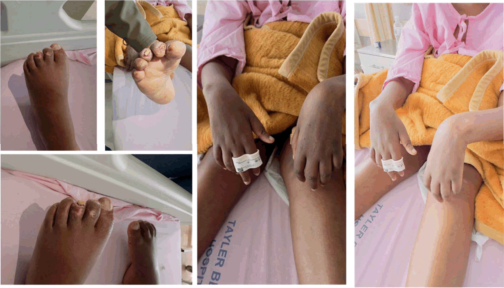
Figure 1: Shows the signs of lupus vasculitis in feet and hands
| Movement | Right | Left |
| Shoulder abduction | 4 | 4+ |
| Elbow flexion | 4+ | 4- |
| Wrist flexion/extension | 2/3 | 0/0 |
| Finger flexion/extension | 3/2 | 0/0 |
| Finger abduction/adduction | 0 | 0 |
| Hip flexion/knee extension | 5/5 | 4+/4+ |
| Ankle dorsiflexion | 0 | 0 |
| Ankle plantarflexion | 4 | 0 |
| Ankle eversion | 1 | 0 |
| Toe extension | 0 | 0 |
Table 2: Motor power assessment (MRC scale, 0–5) at admission
Investigations
Nerve conduction studies and electromyography: Nerve conduction studies performed on admission revealed that of eight sensory nerves tested, three in the proximal upper limbs (bilateral lateral antebrachial cutaneous, left medial antebrachial cutaneous) had normal responses, indicating intact proximal sensory axons. Responses were absent in five nerves: Bilateral median digital (digit II), bilateral ulnar digital (digit V), and the left sural nerve. Distal motor responses in the lower limbs were also absent. Needle EMG showed abnormal spontaneous activity compatible with acute denervation in the left gastrocnemius, left first dorsal interosseous, and left biceps brachii, with reduced interference patterns and altered motor unit morphology. The left tibialis anterior and quadriceps were electrophysiologically normal. The neurophysiology report concluded: Absent distal motor responses with absent SNAPs; an electrophysiological profile showing a proximalto- distal gradient; normal proximal upper-limb studies; EMG evidence of denervation; absence of demyelination; findings consistent with an axonal neuropathy; and a recommendation to exclude ischaemic neuropathy and consider sural nerve biopsy. The lack of demyelinating features is pivotal. The EAN/PNS 2021 criteria for CIDP require across at least two nerves one or more of: Motor conduction velocity <75% of the lower limit of normal; distal motor latency >130% of the upper limit of normal; or conduction block/temporal dispersion. None of these were present. Therefore, CIDP is electrophysiologically excluded, and the axonal, distal-to-proximal gradient aligns with ischaemic injury of the vasa nervorum.
Muscle biopsy: A biopsy of the left hypothenar muscle on 5 February 2026 showed neurogenic atrophy: Fibre size and shape variability, atrophic fibres, increased endomysial fat and collagen, and absence of internal nuclei and regenerating fibres. Focal necrotic fibres were noted. Immunohistochemistry demonstrated sparse CD3-positive T lymphocytes without CD4 or CD8 predominance; CD20 and CD68 stains were negative. The normal serum CK (23 U/L) and negative CD68/CD20 immunostains exclude primary inflammatory and necrotising myopathies — including anti-HMGCR and anti-SRP conditions — on histological grounds. Prior corticosteroid exposure may have dampened inflammatory infiltrates, so the biopsy cannot be assumed to reflect peak inflammatory burden. Lack of fresh tissue prevented enzyme histochemistry and ATPase fibre-typing. Interpreted alongside the NCS, the biopsy confirms a neurogenic cause for the atrophy but does not provide the vasculitic histopathological confirmation that a sural nerve specimen would yield.
Echocardiography: Doppler transthoracic echocardiography showed preserved LV systolic function (EF 65%) and no pericardial effusion. The most striking abnormality was thickening at the tips of the anterior mitral leaflet with preserved valve area and no significant regurgitation, raising the possibility of Libman–Sacks vegetations [9]. Transthoracic imaging has limited sensitivity for this lesion; no transoesophageal study was performed, so this finding should be considered suggestive rather than diagnostic.
Laboratory investigations: Longitudinal and admission laboratory data are summarised in Table 3. Several points deserve emphasis. ESR was elevated in February 2025, rose further on admission, and peaked at 140 mm/hr by discharge, indicating sustained systemic inflammation while CRP remained low throughout — a familiar pattern in active SLE. Haemoglobin decreased from 13.7 to 11.9 g/dL during admission, consistent with anaemia of chronic disease. Platelets fell from 190 to 79 × 109/L by day seven, a clinically significant thrombocytopenia. Complement C3, low in February 2025, had normalised by admission, reflecting fluctuating disease activity. HbA1c (5.5%), vitamin B12, folate, and ACE were normal, excluding diabetes, nutritional deficiencies, and sarcoidosis. HIV, HTLV, and syphilis screens were negative (Table 3).
| Parameter | Feb 2025 | Admission | Discharge | Reference |
| Haemoglobin (g/dL) | 9.1 ↓ | 13.7 | 11.9 ↓ | 12.0-15.0 |
| Platelets (×109/L) | 266 | 190 | 79 ↓ | 150-400 |
| ESR (mm/hr) | 48 ↑ | 43 ↑ | 140 ↑ | <20 |
| CRP (mg/L) | 9 | 5 | 1 | <10 |
| Albumin (g/L) | 30 ↓ | 37 | 32 ↓ | 35-52 |
| Creatinine (µmol/L) | 48 ↓ | >60 | >60 | 49-90 |
| Complement C3 (g/L) | 0.76 ↓ | 1.27 | - | 0.90-1.80 |
| Complement C4 (g/L) | 0.19 | 0.19 | - | 0.10-0.40 |
| Creatine kinase (U/L) | - | 23 | - | 20-180 |
| HbA1c (%) | - | - | 5.5 | <6.5 |
| Vitamin B12 (pmol/L) | - | 355 | - | 156-672 |
| HBV surface Ag / HCV Ab | Neg/Neg | - | - | - |
| HIV/HTLV/RPR | - | All Neg | - | - |
| Urine protein: creatinine (g/mmol, Jun 2025) | - | 0.102 ↑ | - | <0.015 |
Table 3: Longitudinal laboratory parameters
Extended immunological panel
Extended serology (Table 4) revealed markedly positive anti-Ro/SS-A (298 U/mL), with negative anti-La, anti- U1RNP, anti-Sm, and anti-ribosomal P. Loss of Sm and RNP reactivity compared with 2023 likely reflects assay platform differences combined with partial serological modulation from hydroxychloroquine. ANCA, RF, and anti-CCP were all negative, arguing against ANCAassociated or rheumatoid vasculitis. Antiphospholipid testing showed anticardiolipin IgG negative (7 GPL-U/ mL), IgM equivocal (11 MPL-U/mL, below the ≥ 40 cutoff), and anti-β2GPI IgG negative. Lupus anticoagulant screen ratios were weakly positive (1.21 and 1.25), but the confirmatory ratio was 0.96 — below the diagnostic threshold — and the mixing study partially corrected, reducing the likelihood of a true inhibitor. A single weak lupus anticoagulant with equivocal aCL IgM and negative IgG does not satisfy the Miyakis 2006 criteria for definite Antiphospholipid Syndrome (APS) [10]; persistence at 12 weeks is required. Cryoglobulin testing and serum protein electrophoresis with immunofixation remain pending; negative HBV/HCV serology lowers the pretest probability of cryoglobulinaemic vasculitis but does not exclude it.
| Investigation | Result | Value |
| ANA (IFA, HEp-2) | Positive | 1:160, homogeneous |
| Anti-dsDNA | Positive | 51.0 IU/mL |
| Anti-Ro/SS-A | Strongly positive ↑↑ | 298.0 U/mL |
| Anti-La/SS-B, anti-U1RNP, anti-Sm, anti-RibP | All negative | 1.7/1.4/0.7 U/mL |
| Anticardiolipin IgG/IgM/IgA | Neg/Equivocal/Neg | |
| Anti-β2-glycoprotein-I IgG | Negative | 2.6 U/mL |
| Lupus anticoagulant | Weakly positive* | Screen 1.25; confirmatory 0.96 |
| ANCA (c/p-ANCA; anti-PR3/MPO) | All negative | PR3 0.3, MPO 0.3 U/mL |
| RF (IgM)/anti-CCP | Negative/Negative | 1.0 IU/mL / 1.7 U/mL |
| Note: Screen 1.21 (threshold <1.20); normalised ratio 1.25; confirmatory ratio 0.96 (not confirmed). | ||
Table 4: Antiphospholipid and extended immunological panel (16/03/2026)
Results and Discussion
Brief comments on differential diagnosis
Lupus vasculitic neuropathy manifesting as confluent mononeuritis multiplex is the most likely diagnosis. The patient has established SLE with robust multi-antigen serology, objective peripheral ischaemic tissue injury with palpable pulses, and a severe distal-predominant axonal neuropathy lacking demyelinating features. CIDP is ruled out by the EAN/PNS 2021 electrodiagnostic criteria [11]. Antiphospholipid-related vasculopathy remains a plausible co-pathology the clinical picture (digital ischaemia, thrombocytopenia, possible mitral valve abnormality) is consistent with microvascular thrombosis — but serology is below classification thresholds [10]; the 12-week repeat will be decisive. Cryoglobulinaemic vasculitis could present similarly; negative HBV/HCV serology reduces likelihood, but serum cryoglobulins and SPEP/immunofixation are outstanding. Other causes were effectively excluded: diabetic neuropathy (HbA1c 5.5%); nutritional deficiency (normal B12/folate); sarcoid neuropathy (normal ACE); HIV/HTLVassociated neuropathy (seronegative); neurosyphilis (negative RPR/FTA-ABS); ANCA-associated vasculitis (c-ANCA, p-ANCA, anti-PR3, anti-MPO all negative); rheumatoid vasculitis (RF, anti-CCP negative); primary inflammatory or necrotising myopathy (CK 23 U/L; CD68/CD20 negative); and CNS infectious contribution (normal CSF with negative viral PCR and GeneXpert). The thrombocytopenia is most simply explained by immune thrombocytopenia secondary to SLE. Thrombotic microangiopathy was considered less likely given the absence of schistocytes, near-normal bilirubin, and non-elevated LDH. APS-related thrombocytopenia remains possible pending repeat testing. Heparin-induced thrombocytopenia was unlikely because the platelet falls preceded enoxaparin initiation. Hydroxychloroquine-induced thrombocytopenia, though reported, is improbable given three years of prior exposure before the decline.
Brief comments on medical therapy
Therapy targeted three overlapping goals: Suppress the autoimmune driver, provide symptomatic and neuroprotective care, and start rehabilitative measures. Prednisolone was increased from 10 mg to 40 mg daily with a taper plan. It remains to be confirmed from local records whether methylprednisolone pulses preceded the oral escalation. Hydroxychloroquine was continued. The neurology team recommended rituximab as first-line immunosuppression, with cyclophosphamide as an alternative; given the gonadotoxic risk of cyclophosphamide, rituximab is a reasonable option in a young woman desiring fertility preservation despite limited specific evidence for its use in lupus vasculitic neuropathy [12-14]. Because of the digital ischaemia and indeterminate antiphospholipid serology, enoxaparin 40 mg subcutaneously daily was started as prophylactic anticoagulation pending repeat serology. Amitriptyline 25 mg at night was commenced for neuropathic pain.
Gastroprotection, bone protection, and isoniazid/pyridoxine TB chemoprophylaxis were co-prescribed the latter important given planned rituximab in a high TB-burden region.
Physiotherapy began with range-of-motion and bridging exercises. Occupational therapy provided bilateral resting hand splints; foot-drop orthoses were requested.
Brief comments on outcome and follow-up
The patient was discharged after eight days to a regional hospital with rheumatology outpatient follow-up arranged. Neurology follow-up was scheduled at six-monthly intervals, reflecting the slow and often incomplete recovery after severe axonal vasculitic neuropathy axonal regrowth occurs at roughly 1–3 mm per day and functional recovery after prolonged denervation may take months to years. Three pending investigations have major implications:
- The 12-week repeat antiphospholipid panel, which will either confirm APS and necessitate long-term anticoagulation or exclude it.
- Renal biopsy to define the class of lupus nephritis suggested by the June 2025 proteinuria and to guide immunosuppressive intensity.
- Serum cryoglobulins and SPEP/immunofixation to exclude cryoglobulinaemic or paraproteinemic contributions.
A clinical review at 6–8 weeks post-discharge is planned; outcome trajectory data will be incorporated prior to final publication.
Brief comments on the clinical relevance of key autoantibodies
We propose that a principal utility of autoantibodies is their function as serological biomarkers, beyond their roles in monitoring disease activity, contributing to pathogenesis, indicating organ involvement, and informing prognosis. The principal autoantibodies encountered in SLE are outlined in Table 5.
| Autoantibody | Prevalence | Clinical features | Mechanism of action | References |
| Anti-dsDNA | 50–70% | Lupus nephritis; flares; complement consumption | Bind double- stranded DNA forming nephritogenic immune complexes, activate complement, and deposit in glomeruli, triggering inflammation and tissue damage. | Wang X., Xia Y, Anti-double stranded DNA antibodies: Origin, pathogenicity, and targeted therapies, Front Immunol, 10(2019):1667. |
| Anti-Sm | 25–30% | Highly specific for SLE; systemic disease; NPSLE | Target snRNPs interfering with RNA splicing; form immune complexes that activate dendritic cells via TLRs, promoting type I IFN production and systemic autoimmunity; contribute to neurotoxicity via CNS penetration. | Wang X., Xia Y, Anti-double stranded DNA antibodies: Origin, pathogenicity, and targeted therapies, Front Immunol, 10(2019):1667. |
| ANA | >95% | Screening; entry criterion in 2019 EULAR/ACR; non-specific | Bind nuclear antigens activate innate immunity via Fc and TLR signaling. | Choi J., Kim S.T., Craft J, The pathogenesis of systemic lupus erythematosus—An update, Curr Opin Immunol, 24(2012):651-657. |
| Anti-RNP | ~40% | Raynaud’s; arthritis; overlap syndromes | Bind U1-RNP forming immune complexes that activate plasmacytoid dendritic cells via TLR7, enhancing type I IFN production. | Savarese E., Chae O.W., Trowitzsch S., Weber G., Kastner B., et al. U1 small nuclear ribonucleoprotein immune complexes induce type I interferon in plasmacytoid dendritic cells through TLR7, Blood, 107(2006):3229–3234. |
| Anti-La/SSB | 10–15% | Cutaneous lupus; neonatal lupus; hematologic involvement | Bind RNA-associated proteins forming immune complexes that activate Toll-like receptors and type I interferon pathways; mediate immune dysregulation. | Naito R., Ohmura K., Higuchi S., Nakai W., Kohyama M., et al. Positive and negative regulation of the Fcγ receptor-stimulating activity of RNA-containing immune complexes by RNase, JCI Insight, 8(2023):e167799. |
| Anti-Ro/SSA | 30–40% | Cutaneous lupus; photosensiti vity; neonatal lupus; hematologic involvement | Target Ro52/Ro60 ribonucleoprot eins, forming immune complexes that activate dendritic cells via TLRs; cross placenta; bind cardiac tissue. | Naito R., Ohmura K., Higuchi S., Nakai W., Kohyama M., et al. Positive and negative regulation of the Fcγ receptor-stimulating activity of RNA-containing immune complexes by RNase, JCI Insight, 8(2023):e167799. |
| ANuA | 70–90% | Early SLE; lupus nephritis | Bind nucleosome complexes; promote immune complex formation, activate complement, and mediate glomerular deposition and glomerular inflammation. | Toubi E., Shoenfeld Y, Clinical and biological aspects of anti-P-ribosomal protein autoantibodies, Autoimmun Rev, 6(2007):119–125. |
| Anti-ribosomal P | 10–20% | Psychosis; depression; neuropsychi atric lupus | Target ribosomal P proteins; potential CNS penetration, disrupt neuronal function, and trigger neuroinflamma tion via cytokine release and immune complex formation. | Toubi E., Shoenfeld Y, Clinical and biological aspects of anti-P-ribosomal protein autoantibodies, Autoimmun Rev, 6(2007):119-125. |
| Anti-C1q | 15–45% | Lupus nephritis | Bind the collagen-like region of C1q, impair apoptotic cell clearance, activate complement, and promote immune complex-mediated inflammation. | Son M., Diamond B., Santiago-Schwarz F, Fundamental role of C1q in autoimmunity and inflammation, Immunol Res, 63(2015):101–106. |
| Anti-histone | ~30% | Drug-induced lupus | Bind to histone proteins within chromatin forming immune complexes; activate complement. | Elbagir S., Mohammed N.A., Oke V., Larsson A., Nilsson J., et al. Anti-histone and anti-nucleosome rather than anti-dsDNA antibodies associate with IFN- induced biomarkers in Sudanese and Swedish SLE patients, Rheumatology, 64(2025):1170-1178. |
| aPLA (LA, aCL, anti- β2GPI) | 30–40% | Thrombosis; pregnancy loss; antiphospholipid syndrome | Bind phospholipid- bound proteins; activate endothelial cells, platelets, and complement; activate pro-coagulants and autoimmune mechanisms. | Knight J.S., Kanthi Y, Mechanisms of immunothrombosis and vasculopathy in antiphospholipid syndrome, Semin Immunopathol, 44(2022):347-362. |
| Note: aCL : anti-Cardiolipin antibody; ANA: Antinuclear Antibody; ANuA: Anti-Nucleosome Antibody; Anti-C1q: Anti-C1q complement antibody; Anti-dsDNA: Anti-double stranded DNA antibody; Anti-La/SSB: Anti-La/Sjögren’s Syndrome type B antibody; Anti-histone: Anti-histone antibody; aPLA: antiphospholipid Antibodies; Anti-RNP: Anti- Ribonucleoprotein antibody; Anti-Ro/SSA: Anti-Ro/Sjögren’s Syndrome type A antibody; Anti-Sm: Anti-Smith antibody; Anti-ribosomal P: Anti-ribosomal P protein antibody; anti-β2GPI: anti-β2 Glycoprotein I antibody; LA: Lupus Anticoagulant; snRNPs: small nuclear Ribonucleoproteins | ||||
Table 5: Major autoantibodies in Systemic Lupus Erythematosus (SLE): Prevalence, clinical associations, pathogenic mechanisms, and supporting references
Antinuclear Antibodies (ANA) are present in over 95% of SLE patients and are the most sensitive screening marker; in many series ANA precede clinical disease onset [15,16], although lower-level ANA can be found in other connective tissue diseases and even in some healthy individuals. ANA positivity is the entry criterion in the 2019 EULAR/ ACR classification (titer ≥ 1:80) [17]. In SLE, ANA can be subclassified into anti-Extractable Nuclear Antigen (ENA) antibodies, anti-DNA/nucleosome antibodies, and antibodies against DNA- or RNA-associated protein complexes [18]. Anti-double-stranded DNA antibodies are highly specific and present in 50–70% of patients [19]. Anti-RNA binding protein autoantibodies — including anti-RNP, anti-Ro/SSA, anti-Sm, and anti-La/SSB — are reported in approximately 50% of SLE cases and are associated with particular clinical patterns [20]. Anti-Sm antibodies have high specificity but low sensitivity (<20%), occurring in a minority (25–30%) of patients and are more frequent in Asian and African populations; they confer diagnostic specificity but limited sensitivity and have been linked in some reports to neuropsychiatric manifestations [21–24].
This case documents a distinct and severe lupus peripheral neuropathy phenotype defined by three simultaneous elements: Electrophysiological confirmation of severe axonal sensorimotor neuropathy with a distal-predominant gradient; macroscopic digital gangrene despite palpable proximal pulses; and histological evidence of neurogenic atrophy in the most affected territory. Each feature alone is recognised in active SLE; their concurrence in a young patient, with neurophysiology, histology, and vascular findings all pointing to a shared ischaemic microvascular mechanism, justifies reporting. Longitudinal outpatient data add context: Objective evidence of active lupus affecting haematological indices, complement, and renal function was present for more than a year before the neuropathy reached clinical attention, suggesting inadequately controlled multisystem disease rather than an isolated neurologic event.
The neurophysiological pattern requires careful interpretation. The neurophysiologist’s description of ‘electrophysiologically symmetrical with a clear proximal– distal gradient’ refers to the distributional gradient rather than implying identical bilateral amplitudes. Clinical examination demonstrated clear left–right asymmetry (lefthand monoplegia, right-hand partial function), consistent with the multifocal nature of mononeuritis multiplex. As Gwathmey and colleagues note [6], when vasculitic infarction affects many nerve territories extensively, the summed electrophysiological appearance may mimic a symmetric polyneuropathy, while the clinical exam retains asymmetric features reflecting the underlying process. The presence of palpable dorsalis pedis and posterior tibial pulses shifts suspicion away from large-vessel atherosclerotic occlusion toward small-vessel processes inflammatory vasculitis of digital arterioles and vasa nervorum, antiphospholipid-mediated microthrombus’s, or a combination thereof. Antiphospholipid serology in this patient is borderline: A weak lupus anticoagulant not confirmed on the phospholipid-dependent step, equivocal aCL IgM, and negative aCL IgG and anti-β2GPI IgG fall short of the Miyakis 2006 criteria for APS [10]. Nonetheless, the clinical constellation digital ischaemia with palpable pulses, thrombocytopenia, and possible mitral valve abnormality is compatible with a microvascular thrombotic process, and subthreshold serology provides biological plausibility. The 12-week repeat will determine whether confirmed APS alters long-term management.
Sub nephrotic proteinuria of ~0.9 g/g creatinine with bland sediment suggests non-proliferative lupus nephritis (Class II or V) rather than proliferative forms (Class III/IV), although histology is required for definitive classification. Renal involvement supports the case for early immunosuppressive escalation and favours interpreting the findings as a single multisystem immunological flare rather than isolated complications. The renal biopsy result will inform whether rituximab is sufficient as monotherapy or whether agents such as mycophenolate or cyclophosphamide should be added.
Nerve biopsy remains the definitive test for vasculitic neuropathy and was recommended by the neurophysiologist. It was not undertaken during this admission because care was shared between hospitals several hundred kilometres apart, the admitting centre lacked sural nerve biopsy capacity within the admission window, and the clinical team judged that the combination of electrodiagnostic, histological, serological, and clinical data provided an adequate basis for prompt empirical immunosuppression. This pragmatic approach aligns, with qualifications, with the Peripheral Nerve Society guidance on non-systemic vasculitic neuropathy [13]. A skin biopsy from the edge of the ischaemic toe — a feasible alternative with potentially high diagnostic yield for small-vessel vasculitis — remains an option for future histological confirmation.
The demographic context is relevant. SLE in people of African ancestry often follows a more severe course than in those of European ancestry, with higher rates of renal and neurological involvement [12]. Contributing factors include genetic differences in complement regulation and innate immunity, socioeconomic determinants such as delayed diagnosis and limited specialist access, and underrepresentation of African-ancestry patients in clinical trials that inform guidelines. Reports of lupus vasculitic neuropathy from sub-Saharan African tertiary centres are correspondingly scarce; this case adds to that literature.
Lupus Vasculitis (LV) can present in diverse clinical patterns depending on the calibre and anatomical site of affected vessels; when peripheral nerves are involved, several phenotypes occur. In Figure 2 we schematically illustrate the common presentations of peripheral nerve disorders.
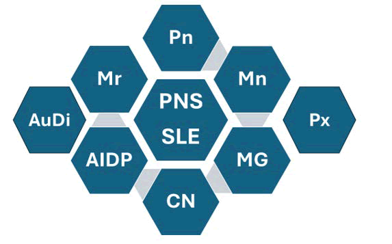
Figure 2: Show a graphical representation of the commonest modalities of peripheral nerves disorder in SLE. Single or multiplex Mononeuritis (Mn), Polyneuropathy (Pn), Monoradiculopathy (Mr), Acute Inflammatory Demyelinating Polyneuropathy (AIDP), Autonomic Disorders (AuDi), Cranial Neuropathies (CN), and Plexopathy (Px).
The most frequent peripheral nerve presentation is mononeuritis multiplex arising from vasa nervorum pathology. The histological components of a peripheral nerve cross-section are depicted in Figure 3.
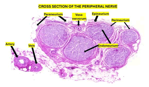
Figure 3: Shows in a cross section of the peripheral nerves their histological components
Additional brief comments on the pathogenesis of lupus vasculitis
The Vasa nervorum (Vn) are small arterioles that supply oxygen and nutrients to peripheral nerves and are essential for normal neurophysiological function. Vn arise from larger arterial branches and form part of the capillary network within the epineurium, perineurium, and endoneurium. Lupus Vasculitis (LV), sometimes termed lupus vasculopathy, affects a substantial proportion of SLE patients presenting with vasculitic features and predominantly involves small vessels, causing organ damage proportional to the vessel calibre and severity, and is generally associated with worse prognosis. Dermatological lesions occur in up to 90% of SLE patients and are frequently accompanied by systemic inflammatory symptoms (fever, weight loss, fatigue) and laboratory abnormalities (raised ESR, anaemia, and other inflammatory markers) as well as Antiphospholipid Syndrome (APS), characterised by aPL positivity including anticardiolipin antibodies, lupus anticoagulant, and/or anti- β2-glycoprotein−1 antibodies [25].
From our review, key pathogenic elements in LV include inflammatory cell infiltration, increased vascular permeability mediated by platelet-derived vasoactive amines and IgE-related mechanisms, and autoantibodies such as anti-endothelial cell antibodies and Antiphospholipid antibodies (APL), as well as ANCA and anti-dsDNA. Immune complexes and chemotactic factors recruit polymorphonuclear leukocytes, leading to endothelial activation with upregulation of adhesion molecules (E-selectin, ICAM-1, VCAM-1) and release of proinflammatory cytokines (IL-1, IL-6, IL-8) and chemokines, resulting in endothelial cell apoptosis and focal vascular wall necrosis. Recruited cells release lysosomal enzymes, including collagenase and elastase, that damage the vessel wall (Figure 4) [25].
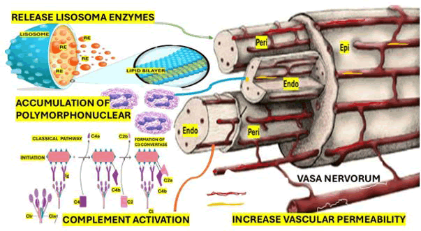
Figure 4: Shows a graphical representation of vasa nervorum. Note: Epi=epineurial vessel, Peri=perineurial vessel, Endo=endoneurial vessels. The extrinsic vessels derive from either arteriae nutriciae or arteria comites and branch into radicular vessels. The intrinsic vessels are supplied by radicular vessels, run longitudinally along the nerve and comprise epineurial, perineurial and endoneurial vessels. Various anastomoses between and within each of these structures arise. It also shows the influence complement activation, lysosome enzymes and polymorphonuclear cells on the pathogenesis on the damage of the vasa nervorum.
Regional increases in vascular permeability, turbulent flow, and hydrostatic pressure at arterial bifurcations promote immune complex deposition, concentrating vascular injury in those sites. Drug-induced vasculitis (from agents such as allopurinol, quinolones, thiazides, pyrazolones, penicillin, retinoids, cytokines, monoclonal antibodies, hydantoins, and various antiepileptics) and infections (e.g., CMV, hepatitis C) can trigger associated vasculitis, sometimes in the context of anti-Ro, aPL positivity, or cryoglobulinemia. Nervous system vasculitis in SLE can affect both central and peripheral compartments [25].
As illustrated in Figure 4, vasa nervorum involvement tends to be focal or multifocal rather than diffuse, producing an asymmetric, progressive, and asynchronous sensorimotor neuropathy involving at least two distinct nerve territories with pain, weakness, and sensory loss, which can sometimes masquerade as a generalized polyneuropathy [26]. Endothelial dysfunction is central to end-organ injury in SLE; impaired endothelial progenitor cell function, blood– nerve interface dysfunction, endothelial activation, and vascular autoantibodies can reduce perfusion to peripheral nerves. In Figure 5 we hypothesise that lysosomal enzyme release, PMN accumulation, and complement activation target the endoneurial microenvironment bounded by endoneurial vessel endothelium and the multilayered perineurium producing local inflammation, vessel occlusion, and necrosis.
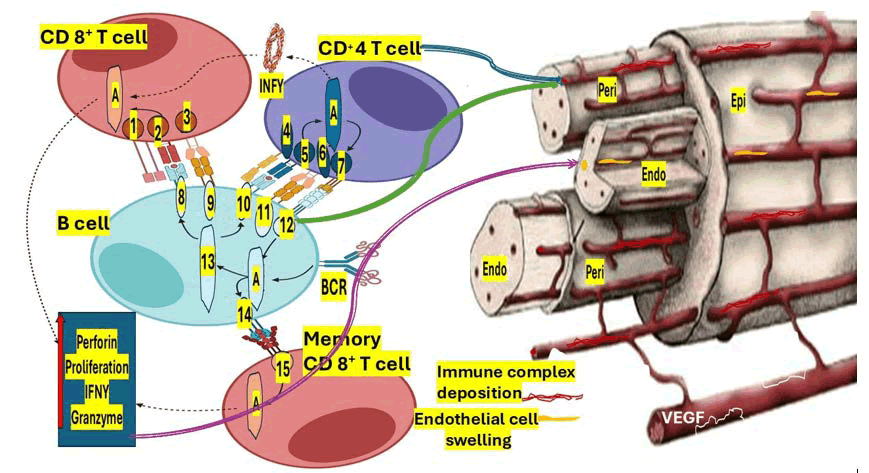
Figure 5: B cells as activators of cellular immunity. Note: B cells are initially activated by antigen recognition through BCR. Internalized antigens are then presented through class II MHC to CD4+ helper T cells, which provide co-stimulatory signals for B cell activation. Activated B cells acquire enhanced potential for antigen presentation with upregulation of MHC-I and II and co-stimulatory molecules, such as CD80/86, further activating both CD4+ and CD8+ T cells. Also, CD27 is upregulated in activated B cells, and interaction between this molecule and CD70 on the membrane of memory CD8+ T cells promotes their maintenance and facilitates their activation in an antigen-independent manner. CD8+ T cell activation leads to efficient cell proliferation and production of potent inflammatory mediators, such as granzymes, perforin, and IFN-γ. 1=CD8, 2=TCR, 3=CD28, 4=CD4, 5=TCR, 6=CD28, 7=CD40L, 8=MHC-I, 9=CD80/86, 10=MHC-II, 11=CD80/86, 12=CD40, 13=Ag Processing, 14=CD27, 15=CD70, VEGF=Vascular endothelial growth factor.
We further speculate that the endothelium and perineurium constrain and regulate exchange between the endoneurial compartment and the surrounding extracellular space, better conceptualised as a blood–nerve exchange and convective endoneurial fluid flow rather than a rigid blood–nerve barrier. We hypothesise that exchange across the interface is governed by hydrostatic pressure gradients that maintain endoneurial homeostasis, and that disruption of these processes compounded in SLE by dysfunctional interactions among immune cells, Schwann cells, axons, tight junctions, and mast cells via altered cell–cell and cell–matrix signalling increases interface permeability and predisposes to mononeuritis multiplex.
Conclusion
Vasculitic ischaemic neuropathy is a distinctive, disabling SLE manifestation that can be diagnosed on electrodiagnostic grounds without nerve biopsy if demyelinating criteria are systematically assessed and excluded. Digital gangrene with palpable proximal pulses in a patient with lupus should prompt evaluation for a shared small-vessel mechanism involving both peripheral nerves and digital arterioles rather than treating these as separate pathologies. Antiphospholipid serology must be performed and interpreted according to formal criteria borderline results are not equivalent to negatives and require confirmatory repeat at 12 weeks because therapeutic implications of confirmed APS are substantial. Longitudinal outpatient testing, when available, enriches clinical interpretation by revealing that multiple organ systems may already be involved sub clinically before neurological deterioration. In resource-limited settings, an integrated clinical synthesis combining neurological examination, electrodiagnostic pattern, peripheral vascular findings, longitudinal serology, and available CSF data can support a robust working diagnosis and justify early aggressive immunosuppression. We reiterate the hypothesis that a major function of autoantibodies is their role as serological biomarkers and we also hypothesised that lysosomal enzyme release, PMN accumulation, and complement activation target specifically on the endoneurial microenvironment bounded by endoneurial vessel endothelium and the multilayered perineurium producing local inflammation, vessel occlusion, and necrosis. We finally hypothesise that exchange across the interface is governed by hydrostatic pressure gradients that maintain endoneurial homeostasis, and that disruption of these processes compounded in SLE by dysfunctional interactions among immune cells, Schwann cells, axons, tight junctions, and mast cells via altered cell–cell and cell– matrix signalling increases interface permeability and predisposes to mononeuritis multiplex.
Learning Points
- Vasculitic ischaemic neuropathy in SLE produces a severe axonal sensorimotor neuropathy with a distalpredominant gradient.
- Formal exclusion of the EAN/PNS 2021 electrodiagnostic criteria for CIDP is the principal discriminator.
- Digital gangrene with palpable proximal pulses in a lupus patient signifies small-vessel microvascular disease.
- Co-occurrence of digital gangrene and axonal neuropathy should be regarded as evidence of a shared ischaemic mechanism rather than coincidence.
- Antiphospholipid serology must be obtained and interpreted against the Miyakis 2006 criteria. Borderline results require confirmatory testing after 12 weeks.
- Longitudinal outpatient laboratory data can be decisive in framing the severity of a flare. Intermittent hypocomplementemia, persistent anaemia, and subnephrotic proteinuria identified during follow-up should prompt escalation of baseline immunosuppression rather than observation.
- Screening before labelling a vasculitic neuropathy in SLE should include ANCA, RF, anti-CCP, hepatitis B and C serology, cryoglobulins, SPEP with immunofixation, HIV, HTLV, syphilis serology, and CSF examination.
- SLE in patients of African ancestry often follows a more aggressive course.
- Electrodiagnostic assessment should be undertaken early in any lupus patient with progressive limb weakness, and the threshold for immunosuppressive escalation should be low.
Authors’ Contributions
All authors participated in the care of the patient, conducted the literature search, and reviewed and approved the manuscript.
Competing Interests
The authors declare no competing interests.
Funding
No specific funding supported this case report.
Patient Consent
Written informed consent for publication of this case and accompanying clinical information was obtained from the patient. A copy is available for review by the Editor-in- Chief on request.
Ethics Approval
This study was not suitable for ethical approval.
References
- ACR Ad Hoc Committee on Neuropsychiatric Lupus Nomenclature, The American College of Rheumatology nomenclature and case definitions for neuropsychiatric lupus syndromes, Arthritis Rheum, 42(1999):599–608.
- J.G. Hanly, Diagnosis and management of neuropsychiatric SLE, Nat Rev Rheumatol, 10(2014):338–347.
- A. Oomatia, H. Fang, M. Petri, J. Birnbaum, Peripheral neuropathies in systemic lupus erythematosus: Clinical features, disease associations, and immunological characteristics evaluated over a twenty-five-year study period, Arthritis Rheumatol, 66(2014):1000–1009.
[Crossref] [Google Scholar] [PubMed]
- B. Florica, E. Aghdassi, J. Su, D.D. Gladman, M.B. Urowitz, et al. Peripheral neuropathy in patients with systemic lupus erythematosus, Semin Arthritis Rheum, 41(2011):203–211.
[Crossref] [Google Scholar] [PubMed]
- G. Said, C. Lacroix, Primary and secondary vasculitic neuropathy, J Neurol, 252(2005):633–641.
[Google Scholar] [PubMed]
- K.G. Gwathmey, T.M. Burns, M.P. Collins, P.J. Dyck, Vasculitic neuropathies, Lancet Neurol, 13(2014):67–82.
[Crossref] [Google Scholar] [PubMed]
- M. Petri, A.M. Orbai, G.S. Alarcón, Derivation and validation of the Systemic Lupus International Collaborating Clinics classification criteria for systemic lupus erythematosus, Arthritis Rheum, 64(2012):2677–2686.
- M. Aringer, K. Costenbader, D. Daikh, European League Against Rheumatism/American College of Rheumatology classification criteria for systemic lupus erythematosus, Ann Rheum Dis, 78(2019):1151–1159.
[Crossref] [Google Scholar] [PubMed]
- I. Moyssakis, M.G. Tektonidou, V.A. Vasilliou, M. Samarkos, V. Votteas, et al. Libman–Sacks endocarditis in systemic lupus erythematosus: Prevalence, associations, and evolution, Am J Med, 120(2007):636–642.
[Crossref] [Google Scholar] [PubMed]
- S. Miyakis, M.D. Lockshin, T. Atsumi, International consensus statement on an update of the classification criteria for definite antiphospholipid syndrome (APS), J Thromb Haemost, 4(2006):295–306.
[Crossref] [Google Scholar] [PubMed]
- P.Y.K. van den Bergh, P.A. van Doorn, R.D.M. Hadden, European Academy of Neurology/Peripheral Nerve Society guideline on diagnosis and treatment of chronic inflammatory demyelinating polyradiculoneuropathy: Report of a joint Task Force Second revision, Eur J Neurol, 28(2021):3556–3583.
[Crossref] [Google Scholar] [PubMed]
- G.J. Pons-Estel, G.S. Alarcón, L. Scofield, L. Reinlib, G.S. Cooper, Understanding the epidemiology and progression of systemic lupus erythematosus, Semin Arthritis Rheum, 39(2010):257–268.
[Crossref] [Google Scholar] [PubMed]
- M.P. Collins, P.J. Dyck, G.S. Gronseth, Peripheral Nerve Society Guideline on the classification, diagnosis, investigation, and immunosuppressive therapy of non-systemic vasculitic neuropathy: Executive summary, J Peripher Nerv Syst, 15(2010):176–184.
[Crossref] [Google Scholar] [PubMed]
- N. Mena-Vázquez, P. Cabezudo García, C. Fuego Varela, S. Manrique-Arija, A. Fernandez-Nebro, Efficacy and safety of rituximab in vasculitic neuropathy: A systematic review of the literature, Reumatol Clin, 15(2019):173–178.
[Crossref] [Google Scholar] [PubMed]
- M. Mahler, P.L. Meroni, X. Bossuyt, M.J. Fritzler, Current concepts and future directions for the assessment of autoantibodies to cellular antigens referred to as anti-nuclear antibodies, J Immunol Res, 2014(2014):315179.
- D.S. Pisetsky, Antinuclear antibody testing misunderstood or misbegotten?, Nat Rev Rheumatol, 13(2017):495–502.
- M. Aringer, K. Costenbader, D. Daikh, R. Brinks, M. Mosca, European league against rheumatism/American College of Rheumatology classification criteria for systemic lupus erythematosus, Arthritis Rheumatol, 71(2019):1400–1412.
[Crossref] [Google Scholar] [PubMed]
- K.H. Ching, P.D. Burbelo, C. Tipton, C. Wei, M. Petri, et al. Two major autoantibody clusters in systemic lupus erythematosus, PLoS One, 7(2012):e32001.
[Crossref] [Google Scholar] [PubMed]
- A. Rahman, Autoantibodies, lupus and the science of sabotage, Rheumatology, 43(2004):1326–1336.
[Crossref] [Google Scholar] [PubMed]
- F. Blanco, J. Kalsi, D.A. Isenberg, Analysis of antibodies to RNA in patients with systemic lupus erythematosus and other autoimmune rheumatic diseases, Clin Exp Immunol, 86(1991):66–70.
[Crossref] [Google Scholar] [PubMed]
- E.J. ter Borg, G. Horst, P.C. Limburg, C.G. Kallenberg, Shifts of anti-Sm-specific antibodies in patients with systemic lupus erythematosus: Analysis by counter-immunoelectrophoresis, immunoblotting and RNA-immunoprecipitation, J Autoimmun, 4(1991):155–164.
[Crossref] [Google Scholar] [PubMed]
- E. Cozzani, M. Drosera, G. Gasparini, A. Parodi, Serology of lupus erythematosus: Correlation between immunopathological features and clinical aspects, Autoimmune Dis, 2014(2014):321359.
[Crossref] [Google Scholar] [PubMed]
- S. Hirohata, Y. Sakuma, T. Yanagida, T. Yoshio, Association of cerebrospinal fluid anti-Sm antibodies with acute confusional state in systemic lupus erythematosus, Arthritis Res Ther, 16(2014):450.
[Crossref] [Google Scholar] [PubMed]
- P. Leone, M. Prete, E. Malerba, A. Bray, N. Susca, et al. Lupus vasculitis: An overview, Biomedicines, 9(2021):1626.
[Crossref] [Google Scholar] [PubMed]
- A. Bortoluzzi, E. Silvagni, F. Furini, M. Piga, M. Govoni, Peripheral nervous system involvement in systemic lupus erythematosus: A review of the evidence, Clin Exp Rheumatol, 37(2019):146–155.
[Google Scholar] [PubMed]
- H.M. Butler, M.E. Zehntner, J.P. van Beusecum, Endothelial dysfunction: Insights into systemic lupus erythematosus-associated cardiovascular disease and neuropsychiatric manifestations, J Cardiovasc Transl Res, 19(2026):36.
Copyright: © 2026 Sibi Joseph, et al. This is an open access article distributed under the terms of the Creative Commons Attribution License, which permits unrestricted use, distribution and reproduction in any medium, provided the original work is properly cited.