

Research Article: Journal of Drug and Alcohol Research (2026) Volume 15, Issue 6
Central Neuropathic Pain from Nociceptive System to Drug Approach: A Comprehensive Review
Sandisiwe Kema1, Lourdes de Fatima Ibanez Valdes2, Sibi Joseph2 and Humberto Foyaca Sibat2*2Department of Internal Medicine and Therapeutics, Nelson Mandela Academic Hospital, Walter Sisulu University, South Africa
Humberto Foyaca Sibat, Department of Internal Medicine and Therapeutics, Nelson Mandela Academic Hospital, Walter Sisulu University, South Africa, Email: humbertofoyacasibat@gmail.com
Received: 05-Feb-2026, Manuscript No. JDAR-26-176235; Editor assigned: 09-Feb-2026, Pre QC No. JDAR-26-176235 (PQ); Reviewed: 23-Feb-2026, QC No. JDAR-26-176235; Revised: 15-Jun-2026, Manuscript No. JDAR-26-176235 (R); Published: 22-Jun-2026, DOI: 10.4303/JDAR/236515
Abstract
Objective: This study aims to review the recent medical literature on therapeutic approaches to central neuropathic pain.
Methods: The systematic review performed in this study followed the guidelines recommended by PRISMA (2020 statement).
Results: A total of 898 records were identified through the database search. Records underwent title and abstract screening, and 752 were excluded. After removal of duplicates, 114 full-text articles were sought for full-text retrieval. 22 records were further excluded because the full text could not be accessed, and 4 were excluded because they could not be fully translated into English. Further studies were excluded because they were irrelevant to central NP. Ultimately, 36 studies were included for this review.
Conclusions: We hypothesised that “affective motivational” and “cognitive evaluative” regions are part of the network involved in emotional and cognitive functions. Among the 36 publications included in this review, none presented a new hypothesis on the neurotransmitters involved in central neuropathic pain. Therefore, to the best of our knowledge, this is the first attempt to graphically represent all the neurotransmitters working on the transmission of neuropathic central pain
Keywords
Introduction
Pain is thought of as a defense mechanism that helps preserve the integrity of possibly damaged tissues by alerting the body to a dangerous stimuli. Emotional, nociceptive, and cognitive problems associated with pain focus attention on the unpleasant input and promote avoidance. Nociceptors (receptors) that receive painful impulses from the skin, muscles, joints, and viscera are classified into four basic types based on the sort of information they send. Both endogenous and external chemical stimuli can activate chemical receptors. Strong mechanical stimulation (high pressure) activates mechanical receptors; extreme temperatures activate thermal receptors; polymodal receptors: Activated by a range of high-intensity stimuli (chemical, mechanical, and thermal) as well as silent receptors, a less common kind of pain receptors that are usually found in the joints and remain dormant to mechanical, chemical, and thermal stimuli. These receptors have the ability to release inflammatory factors brought on by tissue damage, such as arthritis [1,2].
Modulation, transduction, transmission, and perception are the fundamental mechanisms of pain. Modulation is mediated by descending pathways that act directly, allowing the transmission system’s activity to be increased or decreased. Sodium and potassium channels, nociceptors, and Transient Receptor Potential (TRP) channels mediate transduction, which starts with the production of receptors (free nerve endings) in response to tissue-damaging stimuli. Conversely, the nociceptive message is transmitted from peripheral receptors to the central nervous system through ascending routes. Lastly, because perception incorporates a variety of sensory inputs into the subjective knowledge of the triggering event, it is a component of higher brain function, which also includes attention, expectancy, and interpretation [1,2].
At the level of spinal cord dermatomes, all noxious stimuli are identified by specialised primary (or first-order) sensory neurons with their cell bodies located in the Dorsal Root Ganglia (DRG). These neurons are bipolar (or pseudobipolar), with axon projections to both the periphery and the spinal cord, where they synapse in the dorsal horn of the SC with the second-order neuron located in the Laminae of Rexed. The peripheral terminals of these neurons form pain receptors (free nerve endings) that activate a wide variety of receptors, such as G-Protein-Coupled Receptors (GPCRs), and ion channels, such as TRP channels, which are gated by specific noxious stimuli. Following stimulation, the receptor generates depolarizing currents that can start action potentials. These action potentials then move via several transduction fibers (axons) that transport sensory information to the dorsal horn of the SC via the DRG. Based on their width and the presence or absence of a myeline coating, these afferent sensory fibers are divided into the following categories: Primary nociceptive afferents, also referred to as peptidergic and non-peptidergic; these are peptidergic nociceptors that are primarily attached to C fibers (though some Aδ fibers are also included in this group). They can release neuropeptides such as substance P and Calcitonin Gene Related Peptide (CGRP), which can bind to G-protein coupled receptors in the dorsal horn and modulate the transmission of pain. On the other hand, nonpeptidergic neurons may have Aδ or C fibers [3].
Patients with NP have other activity patterns which include relative hypoactivity of the ventromedial and orbitofrontal cortex, which contribute to deficient processing of pain, apart from the emergence of thalamic hypoactivity contralateral to pain stimulation and a shift in pain-related activations from contralateral to ipsilateral posterior insula cortex, which together suggest that additional pathologyspecific reorganisations may contribute to persistent pain, allodynia, and hyperpathia [4].
Across pain aetiologies, consistent changes are also seen in salience and frontoparietal attention networks, including Anterior Cingulate Cortex (ACC), Anterior Insula Cortex (AIC), and medial prefrontal regions. In other words, condition-specific network reorganisation is more often seen in somatosensory regions [5].
A neural pain signature can accurately predict acute pain from distributed brain activity, but performs poorly when applied to clinical chronic pain states [6].
Other authors documented that, in chronic pain, a fundamental reorganisation of brain function, applying new mathematical methods from graph theory to brain networks, found that across pain conditions, chronic pain is related to large-scale disruptions in the overall pattern of brain-wide connections (i.e., network topology) [7].
While other investigators reported that such large-scale disruptions may reflect dysfunction in highly connected network hub regions, leading to a disorganised and hyperconnected brain [8,9].
Vachon-Presseau and collaborators proved that early volumetric changes in the nucleus accumbens and insula predict the transition from subacute to chronic low back pain [10].
Changes in these circuits in chronic pain mirror modifications reported in mood and anxiety disorders and are implicated in most neuropsychiatric disorders [11].
The main aim of this review is to look for an answer to the following research questions:
- What is the latest information on central NP?
- What is the new drug therapeutic approach to central NP?
Materials and Methods
PubMed, Medline, and Cochrane review databases were searched using the term “Central Neuropathic pain” to look for novel clinical reports, diagnostic procedures, and pathogenesis. The study period was filtered from 2020 to 2026. An article type filter was added, and only English articles were reviewed. This systematic review followed PRISMA (2020) guidelines.
To provide an accurate assessment of this search, the corresponding author used a QUADAS-2 evaluation to determine that the risk of bias was low/moderate for almost all publications, and we considered the substantial technical differences observed across diagnostic protocols used in several studies. Notably, in some publications, small and mixed cohorts were analysed, including different types of diagnostic procedures, resulting in fewer cases examined under the same protocol.
Search strategy
From 01 January 2000 to 31 January 2026, we searched the medical literature following PRISMA guidelines. We used these Boolean terms: “Central neuropathic pain” AND “pathogenesis of neuropathic pain” OR immunopathogenesis OR diagnosis OR outcomes) AND (systematic review OR clinical study OR cohort review). We systematically searched the mentioned databases to identify articles on the cited issues.
Only English-language articles were selected. Editorials, letters to the editor, preclinical studies, and conference proceedings were excluded.
Selection of study
The first author screened abstracts and titles, while others independently assessed full texts for eligibility. Publications lacking a clear diagnostic protocol, analysis, complete data, or specifics on patient numbers or AE treatment were excluded.
Selection criteria
Inclusion criteria: Articles with detailed pathogenesis, clinical features, and CNP demographic data.
Exclusion criteria were: (1) Inaccessible full text; (2) Articles not addressing pathogenesis or neurotransmitter for NP; (3) Lack of relevant clinicopathological data; (4) Non-original studies (editorials, letters, conference proceedings, book chapters); (5) non-English publications.
Data extraction and quality assessment
Study quality was rated as good, poor, fair, or reasonable according to NIH and QUADAS-2 criteria. All authors conducted separate quality evaluations, resolving disagreements through discussion and consensus.
Data collection, extraction, and bias assessment
All abstracts and titles meeting the inclusion criteria were reviewed by the first and other authors to collect relevant information for the review. For each selected publication, data on gender, age, publication year, country, study type, total cases, and NP patient treatment were collected. Data from eligible publications were entered into an updated Excel spreadsheet.
Outcome measures
We planned to select the most relevant publications on NP pathophysiology and therapy. This investigation also sought to identify novel theories on the role of neurotransmitters in neural synaptic transmission and the modulation of central NP.
Statistical analysis
Statistical analysis was performed using XLSTAT (add-on for Microsoft Excel, version 2021.4.1, Addinsoft SARL and RStudio.
Results and Discussion
Literature search
A total of 898 records were identified through the database search. Records underwent title and abstract screening, and 752 were excluded. After removal of duplicates, 114 full-text articles were sought for full-text retrieval. 22 records were further excluded because the full text could not be accessed, 52 publications did not address central neuropathic pain, and 4 were excluded because they could not be fully translated into English. Further studies were excluded because they were irrelevant to central NP. Ultimately, 36 studies were included for this review (Figure 1).
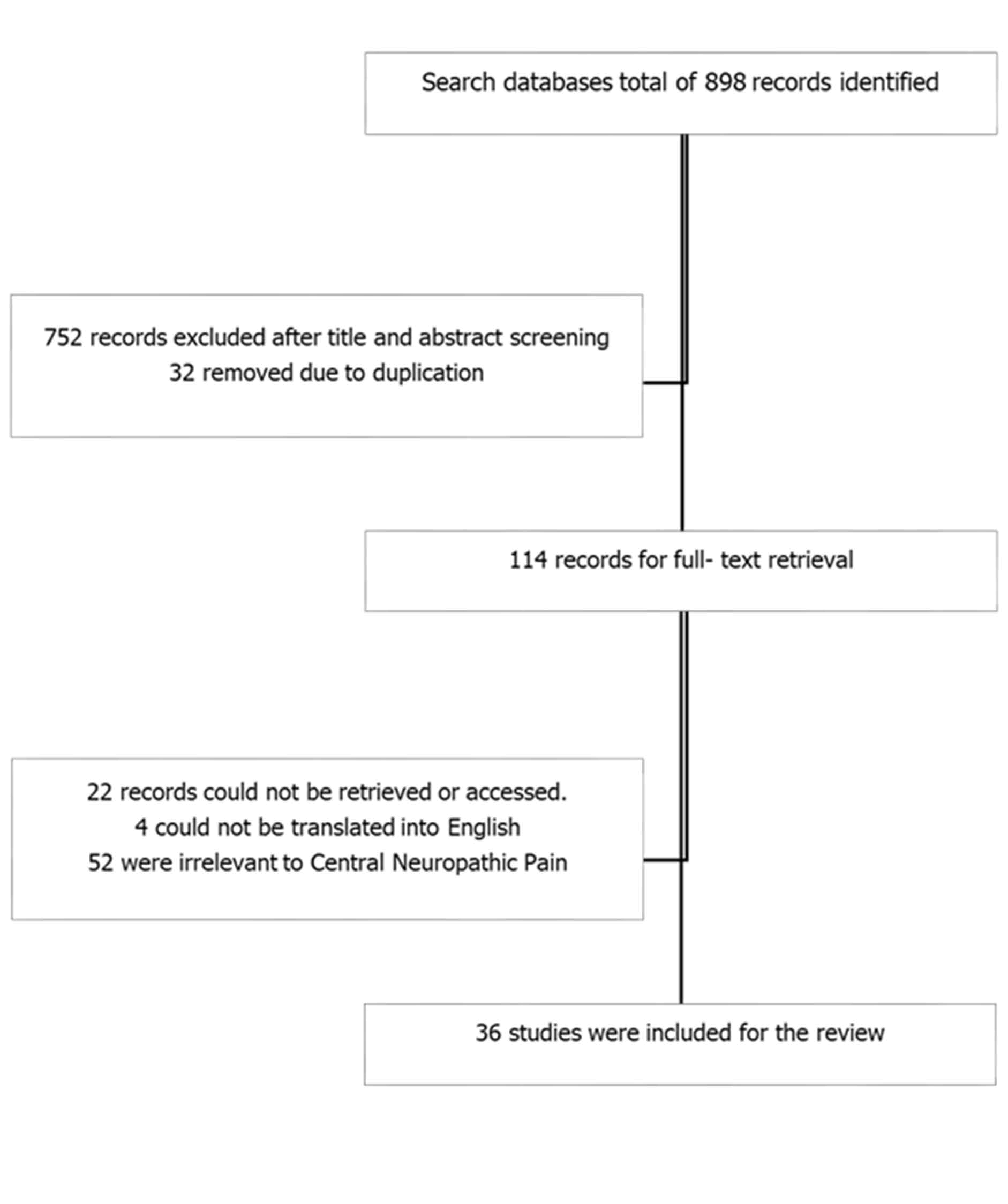
Figure 1: Shows flow diagram of the selected articles
Brief comments on the dorsal horn of SC
All the nociceptive information arriving from the peripheral primary sensory receptor ends at the dorsal horn of the SC. This portion of the SC is divided into ten layers (Laminae of Rexed) according to the cytoarchitectural pattern of the grey matter. Each lamina has a specific functional feature; for example, Lamina I (marginal zone) is responsible primarily for the transduction of the nociceptive information, while Lamina II (substantia gelatinosa) mediates information processing. The cell types within each layer were reflected in this functional stratification: Lamina I has mostly projection neurons and few interneurons, whereas Lamina II has many excitatory (glutaminergic) and inhibitory (GABAergic, glycinergic) interneurons. Each primary afferent in the posterior horn of the SC, such as Aδ´ fibers, ends mostly in Lamina I, with multiple branches synapsing into deeper layers like V and X. However, superficial layers like Lamina I and the outside part of Lamina II are where peptidergic Aδ´ and C afferents terminate. In addition, nonpeptidergic C fibers synapse in the middle region of Lamina II, and both types of fibers (Aδ´ and C) send information about harmless stimuli to Lamina V [11].
The first-order sensory neurons in the DRG send signals to the previously mentioned projection neurons from the dorsal horn (second-order), whose axons decussate to the contralateral side and rise through the anterolateral quadrant of the SC to the thalamus. There are one division of this known spinothalamic tract. the lateral division, which projects to the thalamuss Ventroposterolateral (VPL) nucleus; this enables accurate localization and detection of harmful stimuli. The medial division is made up of neurons that respond to the emotional and cognitive aspects of pain by synapsing in the intralaminar nuclei of the thalamus. These neurons have broad and complicated receptive fields.
The nociceptive, emotional, and cognitive aspects of pain are made possible by the synapse between second-order sensory neurons and third-order sensory neurons that transmit the sensory information to specialized regions after it reaches the thalamic nuclei [11].
The primary Somatosensory cortex (S1), the association and Prefrontal Cortices (PFC), the mid and anterior cingulate cortex, the insular cortex (AI: Anterior Insula; PI: Posterior Insula), the secondary Somatosensory cortex (S2), the amygdala, the Ventral Tegmental Area (VTA), and the Nucleus Accumbens (NAc) are among the cortical regions that are typically involved in processing nociceptive information. However, in addition to the limbic forebrain, hypothalamus, prefrontal cortex, and central nucleus of the amygdala, the descending pathways from the Periaqueductal Grey Matter (PAG) also regulate pain. It is linked to the Rostroventromedial Medulla (RVM), which receives projections from the noradrenergic locus coeruleus and the PAG [11].
Brief comments on the nociceptive system and its functional organisation
For a better understanding of the pathophysiology of Neuropathic Pain (NP) conditions, it is mandatory to understand the neuroanatomy of the nociceptive system and its functional organization, including how its three main compartments (peripheral, spinal cord, and brain) function.
In Table 1, we summarized the most important elements involved in pain definition, fundamental and process before to move forward to other issues.
| Definition | Pain is an unpleasant sensory and emotional experience associated, or resembling that associated, with actual or potential tissue damage. |
| Pain fundamentals | Types of pain based on: The body region involved The system causing the noxious stimuli The duration (acute vs. chronic) |
| The underlying mechanism (somatic, visceral, referred). | |
| Types of receptors (nociceptors): Chemical Mechanical Thermal |
|
| Polymodal | |
| Primary nociceptive afferent fibers: A-delta (Aδ): myelinated, fast fibers |
|
| C: unmyelinated, slow fibers | |
| Key processes | Transduction |
| Transmission | |
| Modulation | |
| Perception |
Table 1: Key facts about the pain pathways
In 2020, some authors defined pain as "an unpleasant sensory and emotional experience associated with, or resembling that associated with, actual or potential tissue damage" [12].
In a few words, the peripheral sensory receptor (free nerve ending) in the epidermis detects noxious stimuli and sends these signals to the posterior horn of the spinal cord, where neuronal circuits process the nociceptive input and transmit ascending nociceptive signals to the brain. Ascending nociceptive information engages complex circuits for top-down neuromodulation and is distributed across brain networks, thereby giving rise to the multidimensional experience of pain [11].
Nociception is the sensory process that identifies harmful stimuli, alerts the body, and protects it from danger through immediate behavioural responses. Sensory pseudo-unipolar neurons found in the Dorsal Root Ganglia (DRG) project to the spinal cord's dorsal horn. Between the periphery and the central neural circuits of the spinal cord, these neurons form a straight "afferent" wire. When painful stimuli are present, the trigeminal ganglion's primary afferent cell bodies target the brainstem trigeminal nuclei. Nociceptive input from the head and face is processed by the nucleus caudalis of the spinal trigeminal nucleus, which is the homolog of the spinal cord dorsal horn. Cell body size and myelination state are the two most important histological characteristics of peripheral sensory neurons grouped together in the DRG. The information from the A-delta myelinated class of nociceptors travels to the spinal cord more quickly than that of C-fibres (free nerve ending/pruriceptors), which makes a significant difference between a first and second pain experience [11].
The dorsal horn of the SC receives, via its interneurons in a laminated architecture, high-threshold nociceptive stimuli and low-threshold, innocuous tactile stimuli through numerous spinal microcircuits that support acute nociception [13].
Itching can be distinguished from pain thanks to these cellular/circuitry mechanisms [14]. However, the brain's interpretation of the activity produced in the spinal dorsal horn projection neurons determines the quality of perception, and inhibitory interneurons are essential to all microcircuit logic in the dorsal horn. However, nociceptive signaling is further expanded by a number of ascending routes via the spinothalamic tract for pain, temperature, and itch, as well as other tracts like spinoreticular, spinoparabrachial, and spinomesencephalic, which engage their respective supraspinal tissues [11].
Molecular subgroupings of spinal projection neurons provide robust evidence of functional divergence within these ascending pathways [15].
The rostral ventral medulla is a crucial component in the pathogenesis of NP, which is represented in Figure 2.
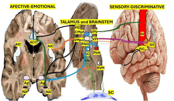
Figure 2: This schematic provides an overarching summary of pain pathways and circuits
Note: Nociceptive information from superficial and deep dorsal horn projection neurons ("Cord") ascends through lateral "sensory" and medial "affective" pathways en-route to brainstem and thalamic relays to specific cortical and subcortical targets. Sensory-discriminative circuits (blue-purple hues, top left) largely originate from deep laminae and ascend through ventral thalamic nuclei (VPM, VPL) to SI. Projections from superficial dorsal horn laminae to VPI thalamus provide a direct route to SII. Both SI and SII project to PIC. Affective-motivational circuits originate primarily from superficial laminae and traverse the parabrachial nucleus and medial thalamic nuclei (MD, CMpf, VMpo) en-route to AIC, ACC, amygdala, and ventral striatum/nucleus accumbens. AIC receives inputs primarily from VMpo, whereas ACC receives inputs from MD and CMpf. Parabrachial nucleus sends overlapping projections to MD, AIC, and ACC, among other regions not shown. Connections between regions and between sensory and affective circuits are extensive but omitted here for clarity
Targeting primary afferent terminals and the cell bodies of second-order neurons that react to painful stimuli, the RVM neurons transmit to the laminae of the dorsal horn of the Spinal Cord (SC) via either attenuating (via inhibition) or amplifying the pain signal, the RVM controls pain in both directions [1,2].
Endogenous opioid peptides, which function as analgesic neurotransmitters, mediate the descending pathways' inhibitory impact. RVM neurons have the ability to activate dorsal horn inhibitory interneurons, which subsequently release opioid peptides including dynorphin and enkephalin. By binding to μ- and k-opioid receptors on afferent terminals, respectively, these peptides reduce neuronal excitability and prevent nociceptive signals from reaching higher levels of the central nervous system. NP and/or inflammatory pain are frequently caused by an imbalance in the PAG-RVM pathway's facilitatory and inhibitory effects. Other members of this network include: 1. Postcentral gyrus; 2. SC posterior horn; 3. Ventral posterolateral nucleus; and 4. Periaqueductal grey material. Figure 2 depicts each of them.
As shown in Figure 2, the expression of the ventral striatum and amygdala responds to noxious input and participates in the early recruitment of descending antinociceptive circuits and to modulate the appropriate behavioural responses to pain [16].
Brief comment on pain modulation circuits
The characteristic of descending projections modulating the spinal nociceptive processing is also represented in Figure 2, including a portion of network-level interactions among brain regions that may modify central pain processing, which include 'cognitive evaluative' regions previously cited. We also hypothesised that the 'pain modulation circuit' comprises regions that modulate pain-related behaviours, such as the expression of pain relief and excitatory stimulation leading to analgesia. The PAG, represented in Figure 2, plays a remarkable role in dorsal horn nociceptive processing and coordinating survival responses to threat, mediated through its top-down influence on arousal, sensory, and motor [17,18].
The PAG projects to the dorsolateral pontine tegmentum and Rostroventral Medulla (RVM) after receiving pertinent inputs from the central nucleus of the amygdala, hypothalamus, ACC, and AIC, among other brain regions. It then projects through the dorsolateral funiculus and specifically targets nociceptive microcircuits in the dorsal horn. Coordinating an opioidergic brainstem antinociceptive circuit is another essential function of PAG. This circuit is broadly projected to the RVM, which contains opposing populations of "ON" and "OFF" neurons that either directly assist or inhibit nociceptive processing at the dorsal horn level. The expression of these "ON" and "OFF" neurons is connected to several state-dependent modifications, such as sleep and arousal [19]. Furthermore, RVM contains spinally projecting serotonergic neurons that do not behave like ON or OFF cells but participate in descending pain modulation. Even though the brainstem contains serotonergic spinal projections, which were initially thought to address descending inhibition, other authors have documented that separable populations of pro and anti-nociceptive dorsal horn-projecting serotonergic neurons in the brainstem with similar monoaminergic projections can have divergent action on spinal nociception [20].
It has been proven that applying opioids over the PAGRVM- Spinal descending pathway inhibits behavioural responses to pain, and local or systemic application of opioid antagonists (e.g., naloxone, naltrexone) blocks the effects of stimulation [21].
As we represented in Figure 2, the most relevant components of the sensorimotor network, are the primary motor cortex, primary and secondary somatosensory cortex; Default Mode Network (DMN) is constituted by the medial prefrontal cortex, posterior cingulate cortex, and angular gyrus; the Frontoparietal Network (FPN) is composed by additional cognitive-evaluative regions such as lateral prefrontal and lateral parietal cortex participating in problem solving, sustained attention, and working memory; and the Salience Network (SN), made by crucial affective regions likes the ACC and AIC, which are which mediate switching among ruminative functions of the DMN and the externally directed attentional functions mediated by the FPN. Regarding pain, intrinsic RSNs clarify how subconscious nociceptive mechanisms gain access to conscious awareness, leading to sustained attention toward pain and the establishment of "remembered pain [22].
Nevertheless, pain attention is related with frontoparietal networks (containing lateral cortical evaluative regions involved in sustained attention) and expression of the salience network (containing ACC and AIC), whereas it’s associated with activation of the default mode network (containing a subset of medial prefrontal and lateral parietal evaluative regions involved in ruminative and introspective processing) [22].
Direct contribution to spinal nociceptive modulation is favoured by connectivity between the PAG and DMN subregions during distraction from pain [23].
It is crucial to remember that deep brain stimulation of the PAG and nearby rostral periventricular grey activates descending antinociceptive pathways; this stimulation increases endogenous opioid release, is reversible by the opioid antagonist naloxone, and is still a viable neurosurgical intervention for the treatment of superrefractory pain conditions [24]. While ACC stimulation alleviates pain escape/avoidance behaviours, PAG lesions block this effect, indicating that the “pain-relieving” effects of ACC stimulation are mediated by the PAG. The ACC is involved in encoding pain aversiveness. Also, ACC cells track pain intensity, and their damage attenuates pain behaviours. Moreover, functional connectivity between ACC and PAG is associated with pain relief and is typically enhanced during successful placebo and opioid analgesia [25,26]. On top of that, projections from the basolateral amygdala to ACC have the opposite action [27].
Furthermore, Tan et al., established that ACC projections through the PIC to the nucleus raphe magnus reinforce peripheral hypersensitivity even when nociceptive input is not certain, leading to another viable pro-nociceptive pathway originating from the ACC.
Nevertheless, rostral subregions of ACC send inputs to the Nucleus Accumbens (NAc), a ventral striatal nucleus involved in reward processing. However, through primarily supraspinal mechanisms ACC-NAc signals promote a hedonic pain-relieving state [28,29].
Subcortical areas, a number of other cortical areas, brainstem locations such as the locus coeruleus (reticular formation), the main source of noradrenergic projections to the brain, the spinal cord, the AIC, and parallel descending pathways are all involved in the modulation of pain. In the sense of opioidergic antinociception, all inputs to the locus coeruleus from RVN and PAG modify its projections to the posterior horn of the SC [30,31].
Brief comments on chronic and NP
A crippling health issue, chronic pain frequently results in drug usage and causes much misery. It is mostly linked to chronic inflammatory processes and damage to the pain pathways, though it may also result from protracted tissue destruction, as is the case with cancer. Trauma, nerve compression, diabetes mellitus, alcohol poisoning, and Herpes varicella-zoster infection are the primary causes of NP, a type of pain that lasts long after the afflicted tissues have recovered [11]. Additionally, NP is linked to peripheral and central sensitization, a kind of plasticity brought on by mediators like chemokines, prostaglandins, and cytokines. Sensitized nociceptors have a lower activation threshold and are located outside the boundaries of damaged areas. Sensitization may therefore make conditions like:
- Allodynia: The perception of pain from harmless stimuli.
- An increased reaction to unpleasant stimuli is known as hyperalgesia.
- Pain without a stimulus is known as spontaneous pain.
Non-Steroidal Anti-Inflammatory Medications (NSAIDs), opiates, antidepressants, and anticonvulsants are the most popular therapy options [11].
Congenital inability for pain perception
Voltage-gated Na+ channels, especially Nav1.7, increase the function of TRP channels in pain perception. The activity of Nav1.7 may be compromised by loss-of-function mutations in the gene encoding one of its component proteins. Despite having intact nociceptive neurons and otherwise adequate somatosensory function, those who are homozygous for this mutation are unable to perceive pain. These people consequently sustain injuries such cuts, bruises, limb fractures, and self-inflicted bites to their lips and mouth. This highlights Nav1.7 as a potential target for the creation of analgesic drugs. The superficial, deep dorsal horn and surrounding white matter (lateral spinal nucleus) exhibit the pertinent projection neurons responsible for transmitting pain stimuli. According to other authors, Aβ- LTMR, low threshold mechanoreceptive afferents that directly connect to deep Lamina V projection neurons, innervate the deep dorsal horn in addition to Lamina I, which is mostly innervated by nociceptors [32].
Strong descending control, mediated by serotonin (5-HT), Norepinephrine (NE), and endorphins that originate in the brainstem, can alter nociceptive processing in several states in all spinal circuits [11].
Specific pathways for sensory and emotional, or affective, pain processing start to diverge in the SC. (e.g., into spinothalamic vs. spinoparabrachial, spinoreticular, and spinomesencephalic tracts) and are later established in the brainstem. The particular information about pain location, intensity and quality is transmitted via the ventral nuclei of the thalamus to the primary and secondary somatosensory cortex at the postcentral gyrus and posterior insular cortex while information related to pain’s inherent unpleasantness is carrying through multiple pathways, mainly via the parabrachial nucleus and medial thalamus to limbic structures, including the ventral striatum, amygdala, anterior insular cortex, and cingulate cortex. The unpleasantness of pain is processed by these limbic structures together. While collateral fibre from these ascending pathways connects with many brainstem nuclei involved in arousal and autonomic functions, further influencing the aversiveness of pain and preparing the organism to mount an appropriate behavioural response (typically escape/avoidance) [11].
As part of the process of parallel ascending projections reaching the brain, it establishes an extensive reciprocal connection with a distributed network of interconnected brain areas.
In pain processing, there is also a well-known pain matrix which projects to higher-order association cortex regions which contribute to a ‘secondary’, or delayed, pain unpleasantness. This process ends in the holistic experience of ‘pain’ (distinct from nociception), and in the selection of a contextually appropriate response, which can range from evolving internal ‘feeling’ states to overt escape behaviours depending on the final brain cortex control [11].
Brief comment on somatosensory pain circuits
The considered ‘sensory-discriminative’ nociceptive processing circuit receives its input from the ascending spinothalamic pathway (‘lateral pain pathway’) via the ventral thalamus to specific cortical targets. This pathway keeps a conserved somatotopic structural composition at each level of the hierarchy. Therefore, neuron cells sampling distinct dermatomes all over the body, including the face, are well-organised with respect to their body site of origin, while axons from the Ventroposterolateral (VPL) thalamus, which receive spinothalamic tract inputs from the body, and Ventroposteromedial (VPM) thalamus from the face and head. The VPL originate mainly in deeper laminae IV and V of the dorsal horn and nucleus caudalis. In turn, the thalamic groups of neurons in the VPL and VPM project to the postcentral gyrus of the parietal lobe, specifically to the primary Somatosensory Cortex (SI), where calculations of stimulus location and intensity take place, via the posterior limb of the internal capsule. As a result, damage to SI hampers the capacity to identify and distinguish noxious stimuli while maintaining affective judgments about how unpleasant they are [33]. Located between the primary Sensory Cortex (SI) and the Posterior Insular Cortex (PIC) in the parietal operculum is the secondary Somatosensory Cortex (SII), where there are higher-order somatosensory neurons able to process nociceptive information, sharing several notable features with VPL/VPM, SI, and inputs primarily from Lamina I spinothalamic neurons of the dorsal horn, including somatotopic responses that correlate with stimulus intensity. It’s an associated cortex with many different functional subregions that receive additional inputs from the limbic system (hippocampus) and parietal lobe, contributing to slightly larger and more complex receptive fields than neurons in SI [34].
The previously cited regions (SII and adjacent PIC) are the only CNS areas shown to elicit painful sensations, with modality, intensity, and location-specific cortical representations of nociceptive stimuli in humans [35,36].
Brief comments on affective pain circuits
The parallel processing of pain unpleasantness is partially mediated by a specific group of dorsal spinal horn projection neurons, initially originating in Lamina I, via the so-called ‘medial pain pathway’ to the brainstem, medial thalamic nuclei, and neocortical ‘affective-motivational’ circuits involved in arousal, autonomic control, and emotional expression. On top of that, other parallel tracts, such as the spinoreticulothalamic pathway (which carries axons from deep lamina VII to the medial thalamus) and direct tracts from the dorsal horn, also send fibres to the basal forebrain, the limbic system (amygdala), and various regions of the neocortex [37].
Conversely, numerous brainstem and diencephalic nuclei, including the medullary and midbrain reticular formations, the locus coeruleus, the Periaqueductal Grey (PAG), the raphe nuclei, and the hypothalamus, are connected to ascending medial pain pathway projections. These connections contribute to early autonomic and arousal changes that facilitate escape readiness. The brain’s “affective-motivational” areas have characteristics that make them ideal for encoding the unpleasantness of pain. Though neurons in affective areas typically have broad, bilateral receptive fields with little somatotopic organization, supporting a more general role in encoding aversiveness, these circuits’ primary function is to modulate the intensity of noxious stimuli, such as “sensorydiscriminative” regions [38].
The main affective brain regions, such as the ventral striatum, anterior insular cortex, amygdala, and anterior cingulate cortex, are broadly implicated in several core processing domains, including learning, reward processing, error monitoring, self-referential processing, and emotional expression, among many more specific functional attributions which are not implicated in somatosensory processing or pain. Nonetheless, unpleasantness is not unique to painful somatic sensations; it is a more general characteristic of noxious and generally aversive stimuli, ranging from non-painful tactile stimulation (e.g., itch) to other salient aversive stimuli across sensory modalities such as auditory, visual, and gustatory. The best investigated cortical regions in the affective circuit are the Anterior Cingulate Cortex (ACC) and Anterior Insular Cortex (AIC) by Positron Emission Tomography (PET) and functional Magnetic Resonance Imaging (fMRI) scans [39].
Damage to the ACC, AIC and the cingulum bundle (white matter tract linking ACC with other pain processing regions)remarkably disrupts the unpleasantness and aversiveness of pain and contributes to an “asymbolia” for pain. Therefore, AIC and ACC are likely required for the experience of pain unpleasantness [40].
The Anterior Insular Cortex (AIC) has an extensive connection with large regions of the cortex, mainly SII and posterior insula and is implicated in various specific functions, including visceral sensation, gustatory processing, and autonomic control and receives projections from the thalamic ventromedial nucleus, posterior part (VMpo); it’s reciprocally connected to the parabrachial nucleus, PAG and is expressed by valence interoceptive stimuli from itch, sensual touch to pain, and bladder distension [41].
The cingulate cortex has a high degree of connectivity along its rostro-caudal axis, with strong connections to limbic and prefrontal structures anteriorly and to premotor structures posteriorly [42].
While, the ACC is a rostral subdivision of the cingulate cortex which participate in a large range of neurophysiological functions such as motor planning, attention, reward processing, cognitive control, memory, decision making, and emotional expression receiving projections from the Mediodorsal (MD) thalamus and adjacent Centromedian parafascicular (CMpf) thalamus (also named as the caudoventral mediodorsal thalamus), which transmits inputs from lamina I of the posterior horn of SC, pontine parabrachial nucleus, and medullary reticular formation [43].
The ACC is involved in several pain-relevant activities and shows crucial cytoarchitectonic and functional differences among its subregions, which are frequently observed in human imaging data and involve more rostral divisions of the midcingulate cortex overlapping with premotor cortex that generate adaptive escape/avoidance responses to threatening stimuli [44].
Some subcortical nuclei such the ventral striatum and amygdala which are strongly related with reward, motivation, emotional expression, and learning are key mediators of pain unpleasantness, being the subpopulation of basolateral amygdala neurons, the one receiving substantial projections from the parabrachial nucleus and projects to descending modulation circuits, including the PAG as it shown while the ventral striatum, (especially the nucleus accumbens), receives extensive dopaminergic projections from the ventral tegmental area, which are essential for reward processing and reinforcement learning [45].
Brief comment on pain circuits
Secondary emotional responses to pain are related to goals, contextual cues, and the integration of somatosensory and affective processing streams with memories, and both generate subjective meanings from the pain experience. The main elements of this ‘affective-motivational’ circuit are ACC and AIC as we before cited plus an extensive reciprocal connections to higher order neurons (that did not tract the pain intensity) such as the orbitofrontal, lateral prefrontal, and lateral parietal structures as have been graphically represented in Figure 3 and which support a ‘cognitive-evaluative’ circuit involved in self-referential and contextual processing activity, relating to the experience of suffering and long-term implications of pain [46,47].
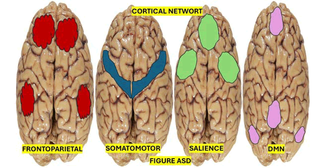
Figure 3: Shows the cortical brain regions involved in central network
These evaluative areas, as mentioned earlier (orbitofrontal cortex/medial prefrontal cortex), are remarkably involved in emotional reappraisal and contribute to the expectation and context-dependent modulation of expression in somatosensory and affective regions, which ‘reinterpret’ or ‘reappraise’ the meaning of a painful stimulus [48].
In a previous publication, we hypothesised that both (“affective motivational”/”cognitive evaluative” regions) constitute a complex and highly interconnected network that participates in emotional and cognitive functions strongly related to the experience of pain and its organisation and specific contributions to acute and chronic pain states [49].
Brief comments on network-level pain processing
The entirety of the brain regions involved in processing pain [50] make up the pain matrix, which accounts for around 15% of the cortical surface and reflects how pervasive pain is across the brain [51]. Despite the ‘affective motivational’ and “cognitive evaluative” circuits being functional attributions to most pain matrix regions, they are not specific to pain. Unfortunately, we still do not know how the holistic experience of pain emerges from the component functions of pain matrix regions accurately; however, based on previous reports, we assumed that the neurophysiological foundation of higher cerebral functions like memory, language and emotion provides a necessary framework to comprehend how complex experiences arise from distributed networks of functionally related brain regions. For example, it’s well known that some injuries in cortical regions may lead to convergent dysfunction in memory or language mechanisms; however, no single “language” or “memory” region is responsible for these processes. Therefore, in each of the previously cited “pain-relevant” brain regions, which were critical to the experience of pain, we might note that inhibiting or activating any one of them would enhance or disrupt pain, while in most cases stimulation of specific brain regions rarely elicits pain and lesions to single regions seldom abolish pain [11].
Based on fMRI findings, other investigators have established how several brain regions interact with complex functions of pain mechanisms, how changes in these connections might influence different pain experiences and how their studies have identified and characterised several intrinsic Resting State Networks (RSNs), which are characterised by synchronised temporal fluctuations in resting brain activity. As shown in Figure 4, the identified RSNs are related to established sensory-discriminative, affective-motivational, and cognitive-evaluative pain circuits [52]. The last one is represented in Figure 4.
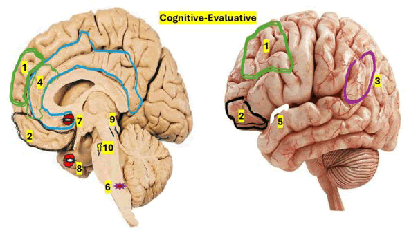
Figure 4: Cognitive-evaluative circuits are not pain-specific but include regions such as: 1) The medial and lateral prefrontal cortex, 2) Orbitofrontal cortex, and 3) Lateral parietal cortex, as well as affective-motivational regions like, 4) Anterior cingulate cortex and 5) Anterior insula cortex. Modulatory circuits include most affective-motivational and cognitive-evaluative regions and other areas, like primary motor cortex, with documented projections to opioidergic and monoaminergic brainstem nuclei that directly modulate incoming nociceptive signals in the dorsal horn. The rostral ventral medulla includes: 6) Nucleus raphe magnus and adjacent reticular formation. There are four canonical Resting State Networks (RSN) linked with pain processing. Other RSNs that are pain relevant, like the limbic network and ventral attention network, are omitted in this picture. Other important components are: 7) Nucleus accumbens, 8) Amygdala, 9) Periaqueductal gray, and 10) Locus coeruleus
Brief comments on brain circuits and networks
Additional changes in motivational behavior, cognitive attentional processes, emotion processing, and memory are present in the majority of chronic pain disorders. These changes combine to produce complex sensations of suffering in pathological chronic pain states. For instance, considerable plastic reorganization of central brain circuits caused by damage to the central nervous system can significantly change their core sensorimotor representations and lead to pathological pain [53].
Central pain syndrome, which is characterized by coexisting sensory disturbance and persistent spontaneous neuropathic pain, is caused by lesions at any level of the spinothalamic tract (including damage affecting terminal projections to the cortex) and is subsequently attributed to plastic reorganization above and below the level of the lesion [53].
The structural changes that occur in a chronic pain setting occur in brain regions containing cognitive-evaluative circuits and affective-motivational circuits. As previously cited, the main cortical regions involved in pain conditions are the AIC, ACC, prefrontal cortex, hippocampus, basal ganglia, SI, and primary motor cortex [54].
The left AIC has been documented as a specific functional marker of chronic pain by Ferraro and colleagues [55].
Brief comments on management and drug therapy
Neuropathic pain is challenging to manage. It is irreversible and at times resistant to treatment. Only 30-40% of patients achieve more than 50% pain relief after treatment. The most relevant drugs administered to central NP are listed in Figure 5 on pharmacological treatment.
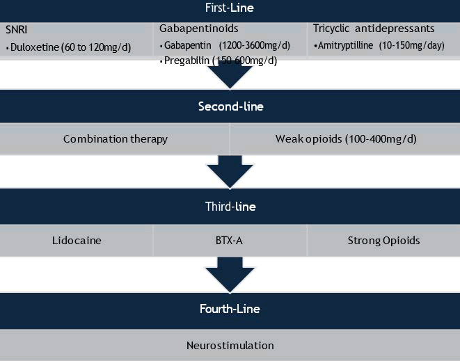
Figure 5: List from the first line to the fourth-line of therapeutic variants
Antidepressants (Tricyclic antidepressants): Within the antidepressant class of drugs, Tricyclic Antidepressants (TCAs) are the most extensively studied and clinically prescribed for neuropathic pain syndromes. Guidelines from professional societies and meta-analyses routinely classify TCAs as a first-line therapy for neuropathic pain [56].
They inhibit reuptake of norepinephrine and serotonin and block synaptic α-adrenergic, serotonergic, histaminic, and muscarinic receptors. Analgesic effects are separable from antidepressant effects and occur at lower doses [57]. Among TCAs, various literature supports amitriptyline as the first-line treatment for neuropathic pain, especially given at a 75 mg per day dose [58].
Dose-dependent anticholinergic effects of TCA included dry mouth, constipation, and urinary retention. Cardiac side effects include orthostatic hypotension and cardiac arrhythmia. TCAs should be used with caution in elderly patients who have had a stroke; they should be thoroughly monitored [58].
Serotonin-Norepinephrine Reuptake Inhibitors (SNRIS): In brain and spinal cord central neurons, duloxetine increases the neurotransmitters serotonin and norepinephrine. In the end, it improves the descending inhibitory pathway’s functionality and rebalances the facilitatory and descending inhibitory systems. Pain perception is inhibited as a result, and the emotional aspects are also modulated. First line for individuals with pain who also have co-occurring anxiety and/or depression [58,59]. Duloxetine has also been shown to inhibit neuroinflammation by decreasing interleukin-8, -12, and interferon-γ [59].
In studies, patients discontinued treatment due to side effects. Side effects include nausea, dizziness, constipation, urinary retention, sexual side effects, hypertension, agitation and somnolence [59-60].
Adverse reactions that can occur include serotonin syndrome and hepatotoxicity [59].
Other SNRIs: There are few trials of other SNRIs in any form of central neuropathic pain. In patients with spinal cord injury, venlafaxine extended release had no effect on neuropathic pain but did improve the intensity of nociceptive pain [57].
Selective Serotonin Reuptake Inhibitors (SSRIs): Limited research is available. From what is available, studies of fluvoxamine showed significant pain relief, and fluoxetine blocked mechanical hypersensitivity and pain, but not thermal-related pain [57,58,61]. Citalopram and trazodone have been reported to be ineffective in treating pain in central neuropathic pain [57].
Anticonvulsants (Gabapentinoids): Largely related to the reduction of dorsal horn sensitivity via binding the α2δ subunit of Voltage-Gated Calcium Channels (VGCC). VGCC consists of subunits (a1 and auxiliary subunits, including α2δ-1 and α2δ-2). The α2δ-1 subunit is expressed in skeletal, cardiac, and smooth muscles, as well as in many neuronal cell types and the Dorsal Root Ganglia (DRG). It is important for behavioural sensitivity and mechanical hypersensitivity. The α2δ-2 subunit is mainly found in the brain, including Purkinje cells in the cerebellum, medulla, hippocampus, and striatum [62].
Overall, gabapentinoids modulate voltage-gated sodium channels, thereby reducing neurotransmitter release. This, therefore, leads to reduced nociceptive signalling. Gabapentinoids significantly improve pain outcomes compared to placebo, with a noted improvement of 30 to 50% from baseline. Adverse effects include somnolence, dizziness and peripheral oedema [63].
Pregabalin is the most researched medication for the management of centralised neuropathic pain; evidence for its effectiveness is mixed [61]. Pregabalin is shown to enhance GABA neurotransmission and reduce glutamate levels; it helps modulate pain, anxiety and insomnia. Pregabalin causes intracortical inhibition in specific neural networks linked to pain and emotional states. It also promotes the production of beta-endorphins and inhibits microglial activation and pro-inflammatory cytokine production [59].
Gabapentin: In a study looking at gabapentin use in patients with spinal cord injury, treatment was found to be effective in reducing pain intensity and frequency >50%, and also noted to improve overall quality of life. Comparison between gabapentin and pregabalin showed that both reduced pains, and no difference in pain relief was found between the two. No studies have compared the adverse-effect profiles of the two [60].
Side effects include weakness, vertigo, sedation, headache and itching [60].
Lamotrigine inhibits glutamate release by selectively binding sodium channels and stabilising presynaptic neuronal membranes. It is the most effective anticonvulsant in the treatment of post-stroke neuropathic pain [61]. In a 2-month trial, lamotrigine showed 44% of patients expressed diminished pain at the highest tested dose of 200 mg PO daily [58].
Side effects and adverse effects: Well-tolerated drug. However, major adverse effects include skin rashes that can lead to life-threatening Stevens-Johnson’s syndrome and toxic epidermal necrolysis [58,60].
Other anticonvulsants: Levetiracetam and carbamazepine have not been shown to be useful in treating post-stroke pain syndrome. Carbamazepine was found to be ineffective in treating post-stroke pain syndrome in a few clinical trials [61]. Phenytoin, lacosamide, sodium valproate, and topiramate are further anticonvulsants that have been suggested for the treatment of central neuropathic pain. More studies are required, nevertheless, as research suggests that there is not enough data to support the use of these medications for neuropathic pain [64].
Opioids neuropathic pain can be effectively treated with tramadol [58]. It possesses SNRI qualities since it is a mild mu opioid agonist that also prevents serotonin and norepinephrine from being reabsorbed [57,58].
Side effects and adverse effects: Constipation, nausea, respiratory depression, reduction in seizure thresholds, serotonin syndrome in combination with 5-HT3 drugs, and a confused state among elderly patients. It does not have dependence potential at therapeutic doses, unlike other opioids like morphine [57,58].
Targeted medication delivery: Treatment-resistant chronic neuropathic pain has been demonstrated to respond well to botulinum neurotoxin A. How it lessens pain is the subject of several theories. Inhibiting glutamate release is one theory [61,64].
One important factor in the regulation of neuropathic pain is glutamate. It has been observed that it is directly linked to the beginning of neuropathic pain and significantly enhances neural excitability after nerve injury. Reduced glutamate release results from BTX-A’s inhibition and downregulation of VGluT2, the vesicular transporter that regulates glutamate storage and release [64]. Additionally, it is believed that BTX-A reduces the expression of pain and inflammatory receptors (TRPV1, P2X3) [57].
Other injections: Although the study only evaluated the effects of lignocaine and baclofen bolus injections within 48 hours, it is unclear how effective they are for long-term pain management [64].
Non-pharmacological treatment (Neurostimulation): Non-invasive brain stimulation/Transcutaneous Electrical Nerve Stimulation (TENS). TENS activates peripheral nerves by delivering electrical pulses to the intact skin surface. It leads to modulation of nerve impulse transmission by inhibiting presynaptic transmission of nociceptive information [65].
Acupuncture-like and intense TENS produce analgesic effects via the descending pain-inhibition system. Normally, when a strong noxious input occurs, it triggers the release of endogenous opioids in the periaqueductal grey and rostral ventral medulla, resulting in descending inhibition of nociception (Endogenous inhibitory control phenomenon). TENS uses this phenomenon [65].
TENS-induced analgesia is also associated with various neurotransmitter systems and their receptors. TENS can increase dynorphin and enkephalin levels in lumbar CSF. Acupuncture-like TENS is associated with increased serotonin release [65].
Clinical application: Conventional TENS delivered for 2 weeks, 8 hours per day, significantly reduced muscle spasm and pain in MS patients; insufficient TENS treatment (only 1 hour per day) did not produce analgesic effects. In a randomised, placebo-controlled trial, TENS had analgesic effects after 6 weeks of treatment (twice a day, 45 minutes each time) [65].
Spinal cord injury: After 2 weeks of treatment (three times per day, 30-40 min per session), conventional TENS and acupuncture-like TENS achieved analgesic effects in 29% and 38% of patients with SC injuries, respectively. The efficacy of acupuncture-like TENS for relieving neuropathic pain in patients with SC injuries was also confirmed across different treatment strategies, e.g., 10 days, 30 min per day, and 12 weeks, 3 times per week, 20 min per time [65].
Central post-stroke pain: Conventional TENS and acupuncture-like TENS applied to the contralateral and/ or ipsilateral sides of the stroke site can relieve pain in a large proportion of patients. TENS has a positive effect on pain reduction and increased mobility in patients with poststroke pain [65].
Repetitive Transcranial Magnetic Stimulation (rTMS) has been widely recommended for the treatment of NP [65]. It is effective, safe and well-tolerated. The analgesic effect is more prominent in patients without depression. The most effective way to deliver rTMS is to target the primary motor cortex with a conventional Figure-8 Coil (F8 Coil) at a relatively high frequency (>5 Hz) [66].
Mechanism of action: rTMS reduces pain by activating the corticospinal and thalamocortical pathways [61]. Additionally, rTMS may lessen the insula, somatosensory, and anterior cingulate cortices’ reactivity to heat and unpleasant stimuli [61]. Additionally, glutamatergic systems, descending pain modulation, and endogenous opioids may be involved in the mechanism of action [66]. However, the exact mechanisms remain unknown [67].
Invasive brain stimulation: Deep Brain Stimulation (DBS) has been used in the management of many neurological diseases, especially essential tremors, other tremors, obsessive-compulsive disorder, neuropathic pain, traumatic brain injury, Tourette’s syndrome, drug-resistant epilepsy and neuropathic pain [61].
The cerebral aqueduct is surrounded by a region of grey matter in the midbrain called the Periaqueductal Grey (PAG). In terms of function, it synchronizes behavioral and autonomic reactions, particularly to pain and other unpleasant stimuli. It incorporates information from the hypothalamus, amygdala, reticular formation, prefrontal cortex, and nociceptive and sympathetic afferents [68,69].
This part of the brain is targeted by DBS for neuropathic pain management, which activates endogenous opioidreleasing neurons that have the ability to block or modify nociceptive signals [68].
52% of patients with neuropathic pain reported goodto- excellent pain reduction (≥ 50% improvement), 23% reported mild relief (20–50% improvement), and 26% reported poor or no effect after stimulation [68]. DBS that targets the PAG has been linked to improvements in hypertension or orthostatic hypotension, as well as benefits in lung function and bladder capacity, in addition to pain management. The implications on autonomic function require more investigation [68]. DBS is more invasive and has a higher risk of serious adverse events. This includes cerebrospinal fluid leak, haematoma, postoperative infection, vertigo, nystagmus and seizures [61].
DBS can be recommended lastly if other therapies are ineffective and must be carefully applied after multidisciplinary consultation [61].
Epidural Motor Cortex Stimulation (EMCS): The EMCS is a surgical intervention that implants an EMCS neurostimulator over the primary motor cortex [70].
Neuroimaging studies have shown changes in brain activity in the thalamus, anterior cingulate cortex, prefrontal cortex, and various regions of the brainstem. However, the exact mechanism of EMCS remains partially unknown [70]. In a few studies on EMCS, it has been shown to provide sufficient pain relief, lasting up to 2 years [61]. EMCS is considered safer than DBS, with the main adverse effects being the risk of postoperative infection, hardware infection and seizures during high-intensity stimulations [61,71].
Other invasive therapies (Neuroablation): Neurosurgery, such as thalamotomy and mesencephalic tractotomy, is a last resort if indicated, and a few reports have shown improvement in allodynia with this surgical intervention. However, complications include normal surgical complications such as infection and haemorrhage, unpleasant adverse effects such as numbness, weakness, or even new-onset neuropathic pain may occur, and the mechanism lacks the adjustability and reversibility of other treatment options [72].
Direct electrical stimulation of the trigeminal ganglion and rootlets using implanted electrodes provided up to 50% pain relief from poststroke facial pain in a small study [61].
Emerging treatments: Mirogabalin is a gabapentinoid, first approved in Japan in 2019 for the treatment of peripheral NP; now, research is looking into treatment for central neuropathic pain. Patients using mirogabalin report improvement in pain from day 6 and an overall improvement in quality of life. Further research is still required [73].
Side effects: Better tolerated than pregabalin; however, side effects include somnolence, dizziness, peripheral oedema, nasopharyngitis, constipation, and weight gain [73].
Ketamine: Given the association of glutamatergic NMDA activity with nociceptive processing, NMDA antagonists, like ketamine, have also been evaluated with some success. However, the cardiovascular and psychological risks associated with the drug class are an area of concern for its potential use. It has been recorded to reduce pain scores among patients with SC injury [72].
Medical cannabinoids: Cannabinoids affect the endocannabinoid system, which is crucial for controlling emotions and senses, as well as the cannabinoid receptors CB1 and CB2. The most prevalent G-protein-coupled receptors in the brain system are cannabinoid receptors. While CB2 receptors are mostly found in peripheral tissues and may be involved in the immune system, CB1 receptors are expressed throughout the brain and central nervous system, including pain pathways in the spinal cord and peripheral nerves [57].
The types of cannabinoids tested for NP conditions include tetrahydrocannabinol and its synthetic analogues dronabinol and nabilone. Cannabinoids require high doses for pain relief; there is also a risk of dependency, and longterm adverse effects are unknown [74].
Potential adverse cognitive and psychiatric outcomes may continue to limit the use of cannabinoids for neuropathic pain in clinical pain [57]. So far, there is no clear evidence for effective pain relief of at least 50% [75].
Non-pharmacological (Stem-cell therapy): Mesenchymal Stem Cells (MSCs) can proliferate and differentiate indefinitely. They exist in adult bone marrow and various tissues, e.g. adipose, nerve and dermis. They can replicate and regenerate as undifferentiated cells indefinitely. They have low immunogenicity and hardly induce the proliferation of allogeneic lymphocytes. They have immunomodulatory, anti-inflammatory and pro-regenerative properties. This is why they have become the highlight of regenerative cell therapy indefinitely [76].
MSCs are set to manage neuropathic pain due to their promotion of angiogenesis, axon regeneration and myelination, neuronal differentiation, the production and secretion of neurotrophic factors, and their immunomodulatory and anti-inflammatory properties indefinitely. More basic research and studies are needed to further explore this treatment modality indefinitely [76].
Vestibular Caloric Stimulation (VCS): The VCS may rebalance the imbalance in bilateral thermal-sensory integration (thermosensory disinhibition hypothesis). This effect was due to the temporary activation of the parietoinsular vestibular cortex. The posterior insula is involved in processing pain, and VCS can activate several areas in the contralateral hemisphere, including the insular cortex [61]. VCS is safe and has shown temporary effectiveness in pain reduction. Repeated CVS can also be recommended if irrigation is tolerated [61].
Psychotherapy: Cognitive-behavioural therapy was useful in the prevention of depression in patients. CBT techniques aim to reframe the patient’s own perception of pain. These techniques address mood, functioning, and social engagement [72].
A recent addition to CBT for the management of chronic pain is Acceptance and Commitment Therapy (ACT). Although it can improve psychological flexibility, it usually does not seem to considerably alleviate pain. Acceptance, awareness, and behavioral adjustments are all components of psychological flexibility. It improves quality of life and helps with depression and general impairment [77].
Psychological relaxation therapy can also be a part of adjunctive treatment [61].
Mindfulness meditation: There is more research on the concept of interoception, or the perception of sensations from within the body. Meditation-mediated stress reduction has been shown to alter brain activity on fMRI and reduce pain in multiple aetiologies of chronic pain [72].
Intestinal microbiota involvement (Probiotic therapy): Probiotics support the growth of normal gut flora, helping maintain intestinal microbiota balance. Research shows that probiotics positively impact gut function by enhancing gut barrier integrity, up-regulating genes involved in mucus secretion, and down-regulating inflammation. Lactobacillus strains F1 and F2 reduce mechanical nociceptive hypersensitivity and cold allodynia, likely by modulating the immune system through expression of TLR2 and TLR4. Further research is necessary; however this should aid in the management of neuropathic pain [78].
Functional microbiota from healthy donors are introduced into the guts of afflicted people through faecal microbiota transplants. By doing this, the balance between Firmicutes and Bacteroidetes in the gut microbiota is restored. Research has indicated promise in the treatment of neuropathic pain. According to research, a fecal transplant can boost the production of 5-HT, further affect the release of neurotransmitters and brain peptides, and quickly produce anti-inflammatory mediators to combat pro-inflammatory processes [78]. This demonstrates NP management potential.
Conclusion
Central neuropathic pain is a complex, multifactorial, and highly disabling condition that arises from lesions or diseases affecting the central somatosensory nervous system. The mechanisms underlying central neuropathic pain involve intricate interactions between neuronal hyperexcitability, dysfunction of descending inhibitory pathways, neuroinflammation and maladaptive neuroplasticity. Emerging evidence also highlights the importance of the corticolimbic system, immunemediated processes, and gut–brain axis interactions in the perpetuation and amplification of chronic pain states.
Despite advances in understanding the neurobiology of pain, the management of central neuropathic pain remains challenging. Future research should focus on identifying precise mechanistic biomarkers, developing targeted therapies, and integrating multidisciplinary biopsychosocial management plans to improve long-term outcomes and quality of life for affected individuals.
We hypothesised that both (“affective motivational” and “cognitive evaluative”) regions are part of the complex, highly interconnected network that participates in emotional and cognitive functions strongly related to the experience of pain, and that this organisation and its specific contributions to acute and chronic pain states.
After injury, disease, or other conditions, changes in anatomy and physiology at any level of the nociceptive system can cause pain, and these changes might contribute to ongoing or sustained pain that outlasts abnormal peripheral nociceptor activity understanding of the relationships between anatomical compartments is crucial to guide the discovery of new drug therapy modalities, which provide the necessary tools for accurate administration of medicines for alleviation or curative purposes. To the best of our knowledge, this is the first review to combine basic science knowledge and therapeutic drugs in central NP.
Acknowledgment
To thanks to Prof Thozama Dubula for his support.
Ethics Statement
This review does not require ethical approval.
Patient Privacy
All patient-identifying information has removed to ensure anonymity.
Conflicts of Interest
Authors of this review report there is not conflicts of interest.
References
- L. de F. Ibañez Valdés, H. Foyaca Sibat, Dercum’s disease associated with autonomic manifestations, novel hypotheses on pathophysiology of neuropathic pain and drug therapy, J Drug Alcohol Res, 14(2025):165079.
- L. de F. Ibañez Valdés, H. Foyaca Sibat, Novel hypotheses on neuropathic pain in Isaacs syndrome and drug therapy, J Drug Alcohol Res, 14(2025).
- S.P. Cohen, L. Vase, W.M. Hooten, Chronic pain: An update on burden, best practices, and new advances, Lancet, 397(2021):2082–2097.
[Crossref] [Google Scholar] [PubMed]
- L. Garcia-Larrea, R. Peyron, Pain matrices and neuropathic pain matrices: A review, Pain, 154(2013):S29.
[Crossref] [Google Scholar] [PubMed]
- F. Cauda, S. Palermo, T. Costa, R. Torta, S. Duca, et al. Gray matter alterations in chronic pain: A network-oriented meta-analytic approach, NeuroImage Clin, 4(2014):676–686.
[Crossref] [Google Scholar] [PubMed]
- D. Lenoir, B. Cagnie, H. Verhelst, R. De Pauw, Graph measure-based connectivity in chronic pain patients: A systematic review, Pain Physician, 22(2021).
[Google Scholar] [PubMed]
- R. De Pauw, H. Aerts, R. Siugzdaite, M. Meeus, I. Coppieters, et al. Hub disruption in patients with chronic neck pain: A graph analytical approach, Pain, 161(2020):729–741.
[Crossref] [Google Scholar] [PubMed]
- J.C. Motzkin, J. Hiser, I. Carroll, R. Wolf, M.K. Baskaya, et al. Human ventromedial prefrontal cortex lesions enhance the effect of expectations on pain perception, Cortex, 166(2023):188–206.
[Crossref] [Google Scholar] [PubMed]
- E. Vachon-Presseau, P. Tétreault, B. Petre, L. Huang, S.E. Berger, et al. Corticolimbic anatomical characteristics predetermine risk for chronic pain, Brain, 139(2016):1958–1970.
[Crossref] [Google Scholar] [PubMed]
- J.C. Motzkin, A.I. Basbaum, A.J. Crowther, Neuroanatomy of the nociceptive system: From nociceptors to brain networks, Int Rev Neurobiol, 179(2024):1–39.
[Crossref] [Google Scholar] [PubMed]
- J.C. Motzkin, A.I. Basbaum, A.J. Crowther, Neuroanatomy of the nociceptive system: From nociceptors to brain networks, Int Rev Neurobiol, 179(2024):1–39.
[Crossref] [Google Scholar] [PubMed]
- S.N., D.B. Carr, M. Cohen, N.B. Finnerup, H. Flor, et al. The revised International Association for the Study of Pain definition of pain: Concepts, challenges and compromises, Pain, 161(2020):1976.
[Crossref] [Google Scholar] [PubMed]
- V.E. Abraira, E.D. Kuehn, A.M. Chirila, M.W. Springel, A.A. Toliver, et al. The cellular and synaptic architecture of the mechanosensory dorsal horn, Cell, 168(2017):295–310.e19.
[Crossref] [Google Scholar] [PubMed]
- X. Dong, X. Dong, Peripheral and central mechanisms of itch, Neuron, 98(2018):482–494.
[Crossref] [Google Scholar] [PubMed]
- R. Wercberger, J.M. Braz, J.A. Weinrich, A.I. Basbaum, Pain and itch processing by subpopulations of molecularly diverse spinal and trigeminal projection neurons, Proc Natl Acad Sci USA, 118(2021):e2105732118.
[Crossref] [Google Scholar] [PubMed]
- N. Schwartz, C. Miller, H.L. Fields, Cortico-accumbens regulation of approach-avoidance behaviour is modified by experience and chronic pain, Cell Rep, 19(2017):1522–1531.
[Crossref] [Google Scholar] [PubMed]
- A.I. Basbaum, H.L. Fields, Endogenous pain control mechanisms: Review and hypothesis, Ann Neurol, 4(1978):451–462.
[Crossref] [Google Scholar] [PubMed]
- S. Koutsikou, R. Apps, B.M. Lumb, Top-down control of spinal sensorimotor circuits essential for survival, J Physiol, 595(2017):4151–4158.
[Crossref] [Google Scholar] [PubMed]
- C.C. De Preter, M.M. Heinricher, The “in’s and out’s” of descending pain modulation from the rostral ventromedial medulla, Trends Neurosci, 47(2024):447–460.
[Crossref] [Google Scholar] [PubMed]
- R.P. Ganley, M.M. De Sousa, K. Werder, T. Öztürk, R. Mendes, et al. Targeted anatomical and functional identification of antinociceptive and pronociceptive serotonergic neurons that project to the spinal dorsal horn, eLife, 12(2023):e78689.
[Crossref] [Google Scholar] [PubMed]
- H. Fields, State-dependent opioid control of pain, Nat Rev Neurosci, 5(2004):565–575.
[Crossref] [Google Scholar] [PubMed]
- L. Garcia-Larrea, R. Peyron, Pain matrices and neuropathic pain matrices: A review, Pain, 154(2013):S29.
[Crossref] [Google Scholar] [PubMed]
- K. Wiech, Deconstructing the sensation of pain: The influence of cognitive processes on pain perception, Science, 354(2016):584–587.
[Crossref] [Google Scholar] [PubMed]
- H. Sims-Williams, J.C. Matthews, P.S. Talbot, S. Love-Jones, J.C. Brooks, et al. Deep brain stimulation of the periaqueductal gray releases endogenous opioids in humans, NeuroImage, 146(2017):833–842.
- T. Schmidt-Wilcke, E. Ichesco, J.P. Hampson, A. Kairys, S. Peltier, et al. Resting state connectivity correlates with drug and placebo response in fibromyalgia patients, NeuroImage Clin, 6(2014):252–261.
[Crossref] [Google Scholar] [PubMed]
- L. Gu, M.L. Uhelski, S. Anand, M. Romero-Ortega, Y.T. Kim, et al. Pain inhibition by optogenetic activation of specific anterior cingulate cortical neurons, PLoS One, 10(2015):1–17.
[Crossref] [Google Scholar] [PubMed]
- K.S. Meda, T. Patel, J.M. Braz, R. Malik, M.L. Turner, et al. Microcircuit mechanisms through which mediodorsal thalamic input to anterior cingulate cortex exacerbates pain-related aversion, Neuron, 102(2019):944–959.e3.
[Crossref] [Google Scholar] [PubMed]
- L.L. Tan, P. Pelzer, C. Heinl, W. Tang, V. Gangadharan, et al. A pathway from midcingulate cortex to posterior insula gates nociceptive hypersensitivity, Nat Neurosci, 20(2017):1591–1601.
[Crossref] [Google Scholar] [PubMed]
- E. Navratilova, J.Y. Xie, A. Okun, C. Qu, N. Eyde, et al. Pain relief produces negative reinforcement through activation of mesolimbic reward-valuation circuitry, Proc Natl Acad Sci USA, 109(2012):20709–20713.
[Crossref] [Google Scholar] [PubMed]
- S.T. Lubejko, G. Livrizzi, S.A. Buczynski, J. Patel, J.C. Yung, et al. Inputs to the locus coeruleus from the periaqueductal gray and rostroventral medulla shape opioid-mediated descending pain modulation, Sci Adv, 10(2024):eadj9581.
[Crossref] [Google Scholar] [PubMed]
- M.W., F. Di Domenico, K. Bannister, A critical brainstem relay for mediation of diffuse noxious inhibitory controls, Brain, 146(2023):2259–2267.
[Crossref] [Google Scholar] [PubMed]
- É. Kókai, W.A. Alsulaiman, A.C. Dickie, A.M. Bell, L. Goffin, et al. Characterisation of deep dorsal horn projection neurons in the spinal cord of the Phox2a::Cre mouse line, Mol Pain, 18(2022).
[Crossref] [Google Scholar] [PubMed]
- M. Ploner, H.-J. Freund, A. Schnitzler, Pain affects without pain sensation in a patient with a postcentral lesion, Pain, 81(1999):211–214.
[Crossref] [Google Scholar] [PubMed]
- S.B. Eickhoff, A. Schleicher, K. Zilles, K. Amunts, The human parietal operculum. I. Cytoarchitectonic mapping of subdivisions, Cereb Cortex, 16(2006):254–267.
[Crossref] [Google Scholar] [PubMed]
- L. Mazzola, J. Isnard, F. Mauguière, Somatosensory and pain responses to stimulation of the second somatosensory area (SII) in humans: A comparison with SI and insular responses, Cereb Cortex, 16(2006):960–968.
[Crossref] [Google Scholar] [PubMed]
- L. Mazzola, J. Isnard, R. Peyron, M. Guénot, F. Mauguière, Somatotopic organization of pain responses to direct electrical stimulation of the human insular cortex, Pain, 146(2009):99–104.
[Crossref] [Google Scholar] [PubMed]
- D.D. Price, Psychological and neural mechanisms of the affective dimension of pain, Science, 288(2000):1769–1772.
[Crossref] [Google Scholar] [PubMed]
- H.L. Fields, Pain: An unpleasant topic, Pain, 82(1999):S61.
- K.L. Casey, S. Minoshima, K.L. Berger, R.A. Koeppe, T.J. Morrow, et al. Positron emission tomographic analysis of cerebral structures activated specifically by repetitive noxious heat stimuli, J Neurophysiol, 71(1994):802–807.
[Crossref] [Google Scholar] [PubMed]
- J.C. Motzkin, J. Hiser, I. Carroll, R. Wolf, M.K. Baskaya, et al. Human ventromedial prefrontal cortex lesions enhance the effect of expectations on pain perception, Cortex, 166(2023):188–206.
[Crossref] [Google Scholar] [PubMed]
- D.F. Cechetto, C.B. Saper, Evidence for a viscerotopic sensory representation in the cortex and thalamus in the rat, J Comp Neurol, 262(1987):27–45.
[Crossref] [Google Scholar] [PubMed]
- A.J. Shackman, T.V. Salomons, H.A. Slagter, A.S. Fox, J.J. Winter, et al. The integration of negative affect, pain and cognitive control in the cingulate cortex, Nat Rev Neurosci, 12(2011):154–167.
[Crossref] [Google Scholar] [PubMed]
- A.D. Craig, J.O. Dostrovsky, Processing of nociceptive information at supraspinal levels, Anesthesia: Biologic Foundations, 21(1997):625–642.
- B.A. Vogt, Pain and emotion interactions in subregions of the cingulate gyrus, Nat Rev Neurosci, 6(2005):533–544.
[Crossref] [Google Scholar] [PubMed]
- S. Ikemoto, Brain reward circuitry beyond the mesolimbic dopamine system: A neurobiological theory, Neurosci Biobehav Rev, 35(2010):129–150.
[Crossref] [Google Scholar] [PubMed]
- M. Roy, D. Shohamy, T.D. Wager, Ventromedial prefrontal-subcortical systems and the generation of affective meaning, Trends Cogn Sci, 16(2012):147–156.
[Crossref] [Google Scholar] [PubMed]
- M. Roy, D. Shohamy, N. Daw, M. Jepma, G.E. Wimmer, et al. Representation of aversive prediction errors in the human periaqueductal gray, Nat Neurosci, 17(2014):1607–1612.
[Crossref] [Google Scholar] [PubMed]
- J. Hiser, M. Koenigs, The multifaceted role of the ventromedial prefrontal cortex in emotion, decision making, social cognition, and psychopathology, Biol Psychiatry, 83(2018):638–647.
[Crossref] [Google Scholar] [PubMed]
- S. Kema, L.F.I. Valdés, S. Joseph, H.F. Sibat, Update information on central neuropathic pain and a new hypothesis on its neurotransmitter, J Drug Alcohol Res, 15(2026).
- R. Melzack, From the gate to the neuromatrix, Pain Suppl, 6(1999):S121–S126.
[Crossref] [Google Scholar] [PubMed]
- M.N. Baliki, A.V. Apkarian, Nociception, pain, negative moods, and behaviour selection, Neuron, 87(2015):474–491.
[Crossref] [Google Scholar] [PubMed]
- J.C. Motzkin, I. Kanungo, M. D'Esposito, P. Shirvalkar, Network targets for therapeutic brain stimulation: Towards personalized therapy for pain, Front Pain Res, 4(2023):1156108.
[Crossref] [Google Scholar] [PubMed]
- J. Rosner, D.C. de Andrade, K.D. Davis, S.M. Gustin, J.L.K. Kramer, et al. Central neuropathic pain, Nat Rev Dis Primers, 9(2023):73.
- R. Kuner, H. Flor, Structural plasticity and reorganisation in chronic pain, Nat Rev Neurosci, 18(2016):20–30.
[Crossref] [Google Scholar] [PubMed]
- S. Ferraro, B. Klugah-Brown, C.R. Tench, S. Yao, A. Nigri, et al. Dysregulated anterior insula reactivity as robust functional biomarker for chronic pain—Meta-analytic evidence from neuroimaging studies, Hum Brain Mapp, 43(2022):998–1010.
[Crossref] [Google Scholar] [PubMed]
- X. Moisset, D. Bouhassira, J. Avez Couturier, H. Alchaar, S. Conradi, et al. Pharmacological and non-pharmacological treatments for neuropathic pain: Systematic review and French recommendations, Rev Neurol, 176(2020):325–352.
[Crossref] [Google Scholar] [PubMed]
- K.N. Gurba, R. Chaudhry, S. Haroutounian, Central neuropathic pain syndromes: Current and emerging pharmacological strategies, CNS Drugs, 36(2022):483–516.
[Crossref] [Google Scholar] [PubMed]
- A.T. Mohanan, S. Nithya, Y. Nomier, D.A. Hassan, A.M. Jali, et al. Stroke-induced central pain: Overview of the mechanisms, management, and emerging targets of central post-stroke pain, Pharmaceuticals (Basel), 16(2023):1103.
[Crossref] [Google Scholar] [PubMed]
- G. Marcianò, M. Evangelista, C. Vocca, V. Rania, C. Palleria, et al. Pregabalin and Duloxetine in patients with non-nociceptive pain: A narrative review exploring the pharmacological effects of this combination, Pharmaceuticals (Basel), 18(2025):1434.
[Crossref] [Google Scholar] [PubMed]
- R.A.A. de Oliveira, A.F. Baptista, K.N. Sá, L.M. Barbosa, O.J.M. do Nascimento, et al. Pharmacological treatment of central neuropathic pain: Consensus of the Brazilian Academy of Neurology, Arq Neuropsiquiatr, 78(2020):741–752.
[Crossref] [Google Scholar] [PubMed]
- S. Ri, The management of poststroke thalamic pain: Update in clinical practice, Diagnostics (Basel), 12(2022):1439.
[Crossref] [Google Scholar] [PubMed]
- F. Yang, Y. Wang, M. Zhang, S. Yu, Mirogabalin as a novel calcium channel α2δ ligand for the treatment of neuropathic pain: A review of clinical update, Front Pharmacol, 15(2024):1491570.
[Crossref] [Google Scholar] [PubMed]
- R.B. João, J.O. Tagore Chan, E.S. Huang, G.P.M. Rosas, A.V. Vasquez, et al. Gabapentinoids for neuropathic pain after spinal cord injury: An updated meta-analysis of randomised controlled trials, Pain Pract, 25(2025):e70060.
[Crossref] [Google Scholar] [PubMed]
- C. Lakra, H. Cohen, A clinical review of the use of Botulinum toxin type A in managing central neuropathic pain in patients with spinal cord injury, J Spinal Cord Med, 45(2022):651–655.
[Crossref] [Google Scholar] [PubMed]
- T. Mokhtari, Q. Ren, N. Li, F. Wang, Y. Bi, et al. Transcutaneous electrical nerve stimulation in relieving neuropathic pain: Basic mechanisms and clinical applications, Curr Pain Headache Rep, 24(2020):14.
[Crossref] [Google Scholar] [PubMed]
- D. Bouhassira, F. Jazat-Poindessous, N. Farnes, C. Franchisseur, A. Stubhaug, et al. Comparison of the analgesic effects of “superficial” and “deep” repetitive transcranial magnetic stimulation in patients with central neuropathic pain: A randomised sham-controlled multicentre international crossover study, Pain, 165(2024):884–892.
[Crossref] [Google Scholar] [PubMed]
- F. Fan, T. Yin, B. Wu, J. Zheng, J. Deng, et al. The role of spinal neurons targeted by corticospinal neurons in central poststroke neuropathic pain, CNS Neurosci Ther, 30(2024):e14813.
[Crossref] [Google Scholar] [PubMed]
- G.J.B. Elias, A. Loh, D. Gwun, A. Pancholi, A. Boutet, et al. Deep brain stimulation of the brainstem, Brain, 144(2020):712–723.
[Crossref] [Google Scholar] [PubMed]
- M.U. Jahngir, A.I. Qureshi, Dejerine-Roussy syndrome, in: StatPearls, StatPearls Publishing, Treasure Island (FL), (2026).
- E. Kurt, R. Witkam, R. van Dongen, K. Vissers, Y. Engels, et al. Preoperative suffering of patients with central neuropathic pain and their expectations prior to motor cortex stimulation: A qualitative study, Healthcare (Basel), 13(2025):1900.
[Crossref] [Google Scholar] [PubMed]
- A. Ramos-Fresnedo, C. Perez-Vega, R.A. Domingo, W.P. Cheshire, E.H. Middlebrooks, et al. Motor cortex stimulation for pain: A narrative review of indications, techniques, and outcomes, Neuromodulation, 25(2022):211–221.
[Crossref] [Google Scholar] [PubMed]
- B.L. Sheldon, Z.T. Olmsted, S. Sabourin, E. Heydari, T.A. Harland, et al. Review of the treatments for central neuropathic pain, Brain Sci, 12(2022):1727.
[Crossref] [Google Scholar] [PubMed]
- T. Ushida, Y. Katayama, Y. Hiasa, M. Nishihara, F. Tajima, et al. mirogabalin for central neuropathic pain after spinal cord injury: A randomised, double-blind, placebo-controlled, phase 3 study in Asia, Neurology, 100(2023):e1193–e1206.
[Crossref] [Google Scholar] [PubMed]
- B.K. Houlind, H.B. Jensen, Medications for managing central neuropathic pain as a result of underlying conditions: A systematic review, Neurol Int, 17(2025).
[Crossref] [Google Scholar] [PubMed]
- G. AteÃ
Â, P. Welsch, P. Klose, T. Phillips, B. Lambers, et al. Cannabis-based medicines for chronic neuropathic pain in adults, Cochrane Database Syst Rev, 3(2018):CD012182.
[Crossref] [Google Scholar] [PubMed]
- W.-J. Zhang, X.-W. Pi, D.-X. Hu, X.-P. Liu, M.-M. Wu, Advances and challenges in cell therapy for neuropathic pain based on mesenchymal stem cells, Front Cell Dev Biol, 13(2025).
[Crossref] [Google Scholar] [PubMed]
- L.M. McCracken, L. Yu, K.E. Vowles, New generation psychological treatments in chronic pain, BMJ, 376(2022):e057212.
[Crossref] [Google Scholar] [PubMed]
- Y. Jiang, H. Xu, W. Zhang, S. Jin, H. Piao, et al. Advances in the study of intestinal microbiota and neuropathic pain, Front Biosci (Landmark Ed), 30(2025).
[Crossref] [Google Scholar] [PubMed]
Copyright: © 2026 Humberto Foyaca Sibat, et al. This is an open access article distributed under the terms of the Creative Commons Attribution License, which permits unrestricted use, distribution and reproduction in any medium, provided the original work is properly cited.