

Case Report: Journal of Drug and Alcohol Research (2026) Volume 15, Issue 3
Bilateral Internal Carotid Artery Aneurysms Presenting as Recurrent Al ternating Oculomotor Palsy: A Case Report with Therapeutic and Drug Considerations
Sibi Joseph1, Saumya Elzebeth Mathew2, Lourdes de Fatima Ibanez Valdes3 and Humberto Foyaca Sibat1*2Department of Neurology, Walter Sisulu University (WSU), South Africa
3Department of Neurology, Nelson Mandela Academic Hospital, Walter Sisulu University, South Africa
Humberto Foyaca Sibat, Department of Neurology, Nelson Mandela Academic Hospital, Walter Sisulu University, South Africa, Email: humbertofoyacasibat@gmail.com
Received: 16-Feb-2026, Manuscript No. JDAR-26-187239; Editor assigned: 18-Feb-2026, Pre QC No. JDAR-26-187239 (PQ); Reviewed: 04-Mar-2026, QC No. JDAR-26-187239; Revised: 17-Mar-2026, Manuscript No. JDAR-26-187239 (R); Published: 24-Mar-2026, DOI: 10.4303/JDAR/236502
Abstract
Alternating oculomotor (cranial nerve III) palsy is a rare symptom of bilateral intracranial aneurysms. Particularly in patients receiving or suspected of exposure to neuroactive or vasoactive drugs, these situations provide a considerable diagnostic difficulty since fluctuating symptoms may be mistaken as neuromuscular diseases.
We describe the case of a 39-year-old HIV-negative lady who had two separate episodes of ophthalmoplegia and unilateral ptosis when she arrived to Nelson Mandela Academic Hospital. The left eye was affected in the first episode in March 2025, and the right eye was affected in December of the same year. Drug-induced neuromuscular junction disorders and medication-related cranial neuropathies were taken into consideration in the differential diagnosis even though the patient had no history of alcohol abuse or recreational drug exposure.
CT angiography showed bilateral saccular aneurysms at the internal carotid artery bifurcations (right: 8 × 4 mm, lobulated; left: 4 × 3 mm), despite a thorough evaluation that produced negative results for autoimmune, infectious, and common neuromuscular etiologies, including the exclusion of drug or toxin-induced causes. Bilateral ophthalmoplegia was established by a thorough ophthalmological evaluation.
This case emphasizes the significance of meticulous medication and substance-use history in clinical diagnosis, as bilateral aneurysmal illness might mimic ocular myasthenia gravis and other drug-related neuromuscular disorders. Additionally, it highlights the diagnostic utility of CT angiography, especially in areas with limited resources where MRI may not be easily accessible. To the best of our knowledge, this is the first instance of recurring alternating ophthalmoplegia and the fourth documented case of bilateral carotid aneurysms at this anatomical position.
Keywords
Introduction
In 1809, the first two documented cases of carotid artery aneurysms were reported [1,2]. We described a 58-year-old woman who had bilateral ophthalmoplegia due to an internal carotid artery aneurysm in the cavernous sinus in 1975. Since then, it has been determined that the most prevalent cause of carotid artery aneurysms, which are bulges of arterial blood vessels supplying blood to the brain and surrounding regions, is atherosclerosis [3]. Significantly, vascular endothelial damage, accelerated atherosclerosis, and an increased risk of aneurysm formation and rupture have all been linked to a number of pharmacological and substance-related risk factors, such as long-term alcohol consumption, tobacco use, and stimulant drugs like cocaine and amphetamines. Vascular wall thinning may also result from long-term usage of corticosteroids and some immunosuppressive medications.
Our patient reported bilateral diplopia, vascular headache, slight eye protrusion on both sides, and facial numbness. Pseudoaneurysm, which only affects one or two layers of the artery wall as a result of local damage, infection, or problems from medical operations, was excluded. Notably, invasive operations and anticoagulant or antiplatelet medication therapy have been linked to iatrogenic pseudoaneurysms, which may increase the risk of bleeding or vascular damage. The first report of a comorbidity between fibromuscular dysplasia and a large aneurysm of the intracavernous carotid artery was made in 1983 by Rebollo and associates [4].
In 1981, Haberbeck-Modesto reported three cases of bilateral aneurysms of the juxta-sellar segment of the internal carotid artery concomitant with intrasellar cisternal herniation, which were misdiagnosed as parasellar tumors [5]. When aneurysms have a diameter exceeding 25 mm, they are defined as giant cerebral aneurysms, which are extremely uncommon [6]. In 20% of patients presenting unruptured intracranial lesions, multiple aneurysms have been reported [6]. In 1999, a pseudoaneurysm of the bifurcation of the internal carotid artery caused by radiation therapy was reported [8].
Pharmacological management plays a critical role in both prevention and supportive care of intracranial aneurysms. Antihypertensive drugs, particularly beta-blockers and calcium channel blockers, are essential in reducing hemodynamic stress on arterial walls. In patients with high risk of rupture, careful use of antiplatelet agents must be balanced against bleeding risk. Moreover, substance abuse, especially chronic alcohol intake, has been associated with poor vascular integrity and increased risk of aneurysm rupture, highlighting the importance of lifestyle modification and addiction management as part of comprehensive care.
When compared to cases with only one aneurysm, series of cases with multiple aneurysms have much worse prognoses and greater fatality rates [9]. An intracranial aneurysm can occur in up to 3% of the general population, and the majority of these cases may not cause any symptoms [10]. According to new research, long-term exposure to alcohol, tobacco, and illegal substances like cocaine and amphetamines raises the risk of aneurysm formation and rupture as well as endothelial dysfunction and hypertension. A 76-year-old woman was described by Gagliardi et al. as having unilateral ptosis that resembled ocular myasthenia and fluctuating bilateral diplopia. A significant bilateral ICA aneurysm in the carotid cavernous tract was confirmed by MRI and magnetic resonance angiography [10]. A 92-year-old female patient with full ophthalmoplegia and right palpebral ptosis was described by Akkari et al. Large bilateral CCAs compressing cranial nerves III, IV, and VI in the right cavernous sinus were confirmed by brain MRI. This case demonstrated an uncommon manifestation of CCA as a potential cause of cavernous sinus syndrome in an older patient who had total ophthalmoplegia [12].
A case of twin large internal carotid aneurysms near the ocular artery that responded favorably to intracranial surgical techniques was reported by Garcia-Bengochea and Deland [13]. Pharmacological techniques are essential for stabilizing patients with intracranial aneurysms in addition to surgical therapy. Antihypertensive medications, such as calcium channel blockers and beta-blockers, are frequently used to lower hemodynamic stress on compromised artery walls and regulate blood pressure. Anticoagulants and antiplatelet medications must also be used carefully because they may raise the risk of hemorrhagic consequences in those who are vulnerable.
When combined with ptosis, diplopia, and pupillary involvement, oculomotor nerve palsy may indicate an underlying compressive aneurysm, making it a high-stakes clinical finding [14,15]. The clinical picture can be further complicated by certain medications, such as sedatives, alcohol, and neuroactive substances, which can temporarily worsen diplopia or mimic neuromuscular junction diseases. Sometimes the diagnosis is difficult to make. Clinicians may first look for neuromuscular, inflammatory, or viral etiologies when clinical symptoms change, go away on their own, or switch between sides; this pattern is known as “ocular pseudo-myasthenia” [3,6]. Vascular imaging and a conclusive diagnosis may be delayed as a result. Less than 1% of all arterial aneurysms and 0.4 to 4% of all peripheral arterial aneurysms are carotid artery aneurysms, making them extremely rare [16]. Crucially, as part of long-term care and prevention programs in at-risk groups, lifestyle modification-such as cutting back on alcohol use and avoiding vasoactive substances should be highlighted.
Third nerve palsy is most frequently linked to intracranial aneurysms that originate from the posterior communicating artery or nearby internal carotid artery segments. The posterior communicating artery (46%) and cavernous internal carotid artery (29%) were shown to be responsible for the majority of oculomotor palsies in a recent systematic study of 513 patients with aneurysm-associated cranial neuropathies [17]. In contrast, it is far less common for bilateral internal carotid artery bifurcation aneurysms to appear with progressive ophthalmoplegia and recurrent alternating ptosis [18,19]. Notably, substance-related factors have been linked to increased hemodynamic stress and arterial wall degradation, which may contribute to the formation of aneurysms in susceptible individuals. These risks include long-term alcohol intake and the use of vasoactive drugs like cocaine. We describe a case where twin ICA bifurcation aneurysms were shown to be the most likely structural reason for a misleading and variable presentation by targeted vascular imaging and persistent clinical suspicion.
Maqsoudi and colleagues reported that a 78-year-old woman had a large thrombosed Intracavernous Carotid Artery Aneurysm (ICCA) exacerbated with ipsilateral carotid thrombosis, a rare and difficult vascular disease. They found that both morphological and hemodynamic factors affect ICCAs, which are rare aneurysms originating from the cavernous section of the ICA. Furthermore, pharmacological factors that affect thrombus development within aneurysms and change the clinical course include long-term use of anticoagulants, antiplatelet medications, and certain hormone therapy. According to the same authors, the presence of thrombus within an intracranial aneurysm raises the likelihood of serious consequences, making accurate imaging evaluation with MRI and MRA necessary to describe the lesion and direct treatment. Furthermore, lowering the risk of aneurysm advancement or rupture requires proper medical management, which includes stringent blood pressure control with antihypertensive medications and close monitoring of drug interactions. They conclude that this study emphasizes the difficulties in diagnosing this illness and the significance of a multidisciplinary approach in choosing the best course of treatment [20].
Case Presentation
A 39-year-old woman was referred to Nelson Mandela Academic Hospital in Mthatha in February 2026 for evaluation of recurrent ptosis. She was HIV-seronegative and had no history of hypertension, diabetes, or prior neurological disease. She was a former smoker (ceased four years prior) and a former alcohol consumer (ceased two years prior).
Her symptoms began in March 2025 with a left-sided headache and left eyelid drooping, which resolved spontaneously after several weeks. Acetylcholine receptor antibody testing performed in April 2025 was negative. She later developed transient diplopia in September 2025, which initially improved.
A second episode occurred on Dec 31, 2025, this time with a right-sided headache, right ptosis, blurred vision, dizziness, photophobia, unsteady gait, speech impairment, facial asymmetry, and hearing disturbance. There was no preceding trauma, fever, or neck stiffness.
On initial assessment, the right pupil was sluggishly reactive to light without frank anisocoria. She had marked right-sided ptosis and severe ophthalmoplegia; vertical and oblique gaze were more restricted than horizontal gaze. The ophthalmoplegia was not painful on examination.
There was no clear fatigability, no bulbar dysfunction, and no additional weakness of the orbicularis oculi. Power was 5/5 in all limbs with normal tone and reflexes. The patient reported gait ataxia and speech impairment, but these were not objectively documented on admission.
Visual acuity was reduced in the right eye (0.3) and preserved in the left (1.0). However, the patient reported persistent subjective visual difficulty in the left eye, which had been affected during the first episode nine months earlier. Classic ophthalmological assessment approximately two weeks’ post-admission confirmed bilateral severe ptosis and ophthalmoplegia: The right eye showed severely restricted movements in all planes (upgaze −4, medial gaze −4, downgaze −4, lateral gaze restricted), while the left eye also showed restriction (upgaze −3, other planes -3). Both pupils had been pharmacologically dilated before this review, precluding assessment of a relative afferent pupillary defect (Figure 1).

Figure 1: Investigations
CT brain with contrast and CT angiography demonstrated bilateral saccular aneurysms at the ICA; the right was lobulated and measured 8 × 4 mm; the left was unilocular and measured 4 × 3 mm (Figures 2 and 3).
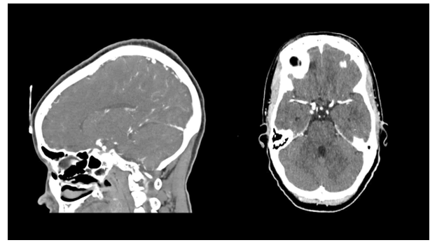
Figure 2: Lateral and axial view of CT scan angiography showing a bilateral aneurysm of the internal carotid artery
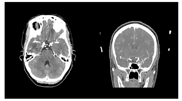
Figure 3: Shows an axial and coronal views of CT angiography showing a bilateral internal carotid artery aneurysm
There was bilateral outward diversion of the globes with structurally normal extraocular muscles. Optic nerves were normal in course and caliber. All other intracranial vessel segments-including the cervical, petrous, cavernous, and communicating segments of both internal carotid arteries, the anterior and posterior communicating arteries, the middle and anterior cerebral arteries, and the posterior circulation-were normal. The graphical representation of the bilateral carotid aneurysm is shown in Figure 4.
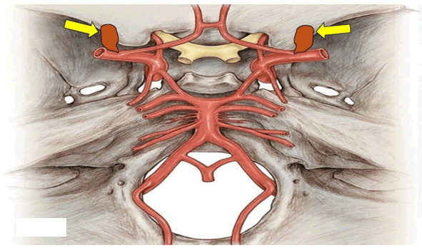
Figure 4: A yellow arrow pointing to the graphical representation of the bilateral aneurysm of the ICA at the bifurcation
There was no evidence of haemorrhage, thrombosis, atherosclerosis, leptomeningeal enhancement, retrobulbar mass, or other focal intracranial lesion.
Further investigations were largely revealed. Autoimmune markers (ANA, anti-PR3, anti-MPO, anti-CCP, HLA-B27) were all negative. Thyroid function tests and TSH receptor antibodies were normal. Cerebrospinal fluid obtained by lumbar puncture on Mar 4, 2026, was clear and colorless with no cells, normal protein (0.23 g/L) and glucose (3.0 mmol/L), absent oligoclonal bands, normal IgG, negative adenosine deaminase, and negative microbiology, including GeneXpert MTB/RIF, bacterial culture, and cryptococcal antigen. CSF VDRL was non-reactive.
Infectious screening (HIV on two occasions, Toxoplasma IgG/IgM, cysticercosis ELISA, cryptococcal antigen) was negative. Syphilis serology showed reactive treponemal antibodies (confirmed by positive FTA-ABS) with a nonreactive RPR on multiple occasions. Hematological and biochemical parameters were largely normal, except for a mild normocytic hypochromic anemia (Hb 11.2 g/dL), low serum iron and transferring saturation consistent with iron deficiency, and an isolated elevation of GGT (123 U/L) attributed to prior alcohol use. HbA1c was normal (5.1%).
MRI could not be performed as it was unavailable at our institution. Anti-NMDA receptor antibodies and aquaporin-4 IgG were referred to a distant centre, and results were pending at the time of writing. The patient remained clinically stable but functionally impaired. Because local neurosurgical expertise for complex vascular lesions was unavailable, she was referred to Inkhosi Albert Luthuli Central Hospital in Durban, South Africa for definitive endovascular or surgical management.
Results and Discussion
The main diagnostic trap in this case was the alternating nature of the symptoms. Recurrent unilateral ptosis and diplopia, especially when symptoms fluctuate over time, naturally raise the possibility of ocular myasthenia gravis [16]. That was a reasonable early consideration here.
However, the presence of mydriasis, the absence of diurnal fatigability, lack of bulbar involvement, negative acetylcholine receptor antibodies, and progressive ophthalmoplegia all ruled out myasthenia as a differential diagnosis.
A similar pattern has been described in bilateral cavernous carotid aneurysms, as we reported before [3], in which fluctuating ocular symptoms initially led clinicians to consider ocular myasthenia before the structural lesion became apparent [18,19]. The term ‘pseudo-myasthenia’ has been used to describe weakness and fatigability of the lids and extraocular muscles caused by cranial nerve compression rather than a neuromuscular transmission disorder [6]. This phenomenon has been reported with posterior communicating artery, basilar artery, and carotid aneurysms [16,18,21] and in one striking case, even a positive edrophonium (Tensilon) test did not exclude aneurysmal compression [21].
The right pupil was sluggishly reactive to light without frank anisocoria, suggesting partial involvement of the parasympathetic fibers of the oculomotor nerve. These fibers run superficially within the nerve and are particularly vulnerable to external compression, though incomplete involvement can occur with partial or intermittent compression [22,23].
Oculomotor palsy is most classically associated with posterior communicating artery, cavernous, and clinoid segment aneurysms [4]. Third nerve palsy arising from an aneurysm at the ICA bifurcation itself is far less commonly described. The anatomical relationship between the ICA aneurysm and other blood vessels and other structures are represented in Figure 5.

Figure 5: Shows the anatomical components around the carotid internal artery aneurysm
However, the terminal segment of the internal carotid artery-C7 in the Bouthillier classification [9] passes between the optic and oculomotor nerves before dividing into the anterior and middle cerebral arteries. A saccular aneurysm arising at this bifurcation point, therefore, sits in direct anatomical proximity to the cisternal segment of the oculomotor nerve, where it runs between the posterior cerebral and superior cerebellar arteries [29,25].
The differences in shape of the saccular aneurysm (A) compared with the fusiform aneurysm (B) are graphically represented in Figure 6.
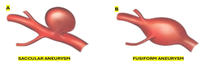
Figure 6: Shows the morphological differences between saccular and fusiform aneurysm. Micotic aneurysm was not considered in this study
CT imaging confirmed bilateral outward diversion of the globes with structurally normal extraocular muscles – a finding consistent with bilateral oculomotor underactivity rather than an orbital process – and formal ophthalmological assessment documented bilateral ophthalmoplegia with more severe restriction on the right, the side of the larger aneurysm. Taken together, imaging and clinical findings support a compressive mechanism, even though the exact point of nerve contact cannot be confirmed without higherresolution vascular imaging [23,24].
In Figure 7, we represent the different types of aneurysms according to their location along to the ICA and the common carotid artery.
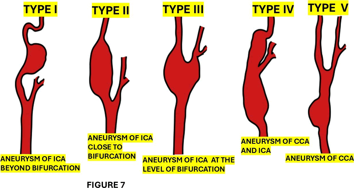
Figure 7: Represent the topographic classification of carotid aneurysm. The graphical representation is self-explanatory
In summary, ICA aneurysms can present with diverse clinical features depending on the affected segment of the artery and the compressive effect of bilateral giant vascular malformations, which require individualized management of the nearby structures in addition to the aneurysm itself [25]. Although CT angiography is less sensitive than Digital Subtraction Angiography (DSA), which remains the gold standard, many authors agree that CTA is highly accurate in diagnosing intracranial aneurysms. Therefore, it can be used as an alternative diagnostic tool rather than DSAN [26,26].
Brief comments on bilateral supraclinoid carotid aneurysm
Supraclinoid aneurysms are in the intradural space, and they originate from the ICA distal to the distal dural ring, to the carotid terminus. Reports of bilateral giant supraclinoid aneurysms are extremely uncommon [28].
Only on two occasions have bilateral supraclinoid aneurysms been reported; therefore, they are extremely rare (Table 1).
| Authors and citation | Age | Gender | Clinical presentation | Examination | Labs | Associated diseases | MRI |
| Badrawi N, Iqbal SS, Ahmed A, Iqbal SS | 44 | F | Chronic headache associated with progressive loss of visual acuity for 6 months. The headache was unilateral to the Lt side and throbbing in nature. The headache was associated with blurring of vision and photophobia. | Reduced visual acuity. | N/A | Bilateral supraclinoid saccular aneurysms identified. The Rt aneur abutted the Rt optic n after the ophtha artery's origin, while the aneurysm compressed Lt optic nerve and chiasm near the ophthalmic artery's or TOF images illustrated these aneurysms. | |
| García Carreira MC, Cánovas Vergé D, Marco Igual M, Hervàs Pujol M | 49 | F | Progressive loss of visual acuity over 5 years. Also had retro- orbital pain for few weeks. | Bilateral papillary pallor and decreased visual acuity. (Rt Eye: 0.8; Lt Eye: 0.5) | Negative or normal | None | Carotid arteries dilation the supraclinoid segm resulting in compression of the o nerves. MR angiogr showed bila aneurysms at emergence of the ophthalmic artery ( 16 × 12 mm, neck 8.38 and the left ophtha artery ( 17 mm, neck mm). |
| Badrawi N, Iqbal SS, Ahmed A, Iqbal SS | 42 | M | Persistent vascular headache and progressive visual loss over time. | Reduced visual acuity | N/A | Hydrocep halus | Brain CT, CT angiogram MRI, and MR angiogr confirmed bilateral la partly thromb supraclinoid seg fusiform aneurysms of ICA compressing. |
| Note: M: Male; F: Female; NA: Not Available or Not Applicable; CT: Computed Tomography; MRA: Magnetic Resonance Angiography; ICA: Internal Carotid Artery; cm: centimeters; mm: millimeters; DSA: Digital Subtraction Angiography; TOF: Time-of-Flight (MRI sequence); Rt: Right; Lt: Left; F/U: Follow Up. | |||||||
Table 1: Literature review findings of bilateral supraclinoid ICA aneurysms
This malformation can cause a wide range of symptoms and require careful management. Khattab et al. reported a 42-year-old man who presented with a persistent vascular headache and progressive visual loss over time. Brain CT, CT angiography, MRI, and MR angiography confirmed bilateral large, partly thrombosed supraclinoid segment fusiform aneurysms of the ICA compressing the midbrain, leading to obstructive hydrocephalus. Their patient denied both surgery and endovascular therapy, but a ventriculoperitoneal shunt was performed, which relieved the patient’s symptoms. Concluding that, bilateral giant aneurysms of the ICA present unique challenges and can lead to various clinical manifestations and effects on surrounding structures [28].
Khattab and colleagues reported a 42-year-old Middle Eastern male patient with the largest bilateral supraclinoid internal carotid aneurysms presenting with hydrocephalus [28].
The bilateral nature of the aneurysmal disease also offers a plausible explanation for the sequential, side-switching pattern of presentation over time [18,21,25]. The negative work-up for common mimics is an important part of the argument. CSF was normal and acellular, with negative microbiology and absent oligoclonal bands. Autoimmune and thyroid testing were unrevealing.
Treponemal serology was reactive, but the RPR and CSF VDRL were non-reactive. CSF was acellular, with normal protein levels, and no oligoclonal bands were detected. This pattern is most in keeping with a Sero fast state from prior treponemal exposure rather than active neurosyphilis. There were also no features of large-vessel vasculopathy on CT angiography – such as arterial wall irregularity, beading, or stenosis – to suggest syphilitic arteritis as a contributor to aneurysm formation. None of this proves that the aneurysms caused the full clinical picture, but it does narrow the differential considerably and makes the vascular explanation harder to dismiss. Upon searching the medical literature, we only found three publications regarding a bilateral supraclinoid carotid artery aneurysm [24,29,30]
Brief comments on treatment for intracranial aneurysms
Therapy for intracranial aneurysms includes reconstructive and occlusive strategies. All occlusive approaches include surgical or endovascular ligation of the parent artery. Reconstructive procedure may include the application of coil embolization with or without a vascular reconstruction device, microsurgical clips, the use of flow-diverting devices, or the employment of liquid embolic agents [31,32]. The graphical representation of the location of the clips at the neck of saccular aneurysm of the bifurcation of the ICA is shown in Figure 8.
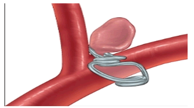
Figure 8: Graphical representation of clipping the neck of saccular aneurysm at the bifurcation of the ICA
As a general procedure, occlusive strategies is used if we cannot preserve the parent vessel which can be achieved by Hunterian techniques involving occlusion of the ICA with or without revascularization which can be performed by 1) Occlusion alone, 2) Occlusion with a low-flow bypass (extracranial artery), and 3) Occlusion with a high-flow bypass (saphenous vein or radial artery) [33]. The microsurgical treatment of aneurysms in the paraclinoid area is a neurosurgical challenge due to the intricate anatomy surrounding the ICA at that level, as well as difficulty in obtaining proximal control and in clinoid drilling, which may significantly impede an accurate clip placement, making endovascular treatment a better choice.
Limitations
Anti-MuSK antibodies and electrophysiological studies (repetitive nerve stimulation, single-fibre EMG) were not performed; seronegative myasthenia gravis, therefore, cannot be completely excluded, though the progressive course, pupillary involvement, and bilateral ophthalmoplegia without fatigability make it less likely. Anti-NMDA receptor antibodies and aquaporin-4 IgG results were pending at the time of writing; neuromyelitis optica spectrum disorder and autoimmune encephalitis have not been formally excluded. MRI and catheter angiography were not available locally, so the precise relationship between the aneurysm wall and the oculomotor nerve could not be confirmed.
This case also reflects the practical challenges of working in a resource-limited setting. MRI and catheter angiography were not available locally, yet careful clinical observation combined with CT angiography was still sufficient to identify a clinically important vascular lesion and redirect management toward urgent neurosurgical referral. CT angiography remains a valuable and accessible first-line investigation when history and examination raise concern for a compressive lesion [27,34,35]. Its sensitivity for intracranial aneurysms has been evaluated in the South African setting, where it serves as a screening tool before digital subtraction angiography in centres where the latter is available [27],34]]. Carotid aneurysms leading to third nerve palsy are often discussed in ideal diagnostic settings, but many clinicians must make decisions with far fewer tools. In that context, clinical vigilance and targeted CTA can be lifesaving [36,37].
Learning points
Do not assume recurrent or alternating ptosis is always myasthenia gravis, especially when there is pupillary involvement or progressive ophthalmoplegia without fatigability.
A sluggishly reactive pupil in the setting of oculomotor palsy should raise concern for a compressive lesion and prompt urgent vascular imaging, even when the broader presentation appears mixed.
Bilateral intracranial aneurysms may present with sequential or alternating unilateral symptoms over time and may convincingly mimic neuromuscular disease.
In settings where MRI is unavailable, CT angiography remains a useful tool for identifying clinically important aneurysmal lesions and guiding timely referrals.
Conclusion
This case describes an uncommon presentation of bilateral internal carotid artery bifurcation aneurysms in a young woman with recurrent alternating ptosis, pupillary involvement, and progressive bilateral ophthalmoplegia. The diagnosis was not obvious initially and became clear only after broader differential diagnoses had been explored and vascular imaging was performed.
Although the exact mechanism of nerve involvement cannot be definitively established without higherresolution imaging, the bilateral aneurysms were the only major structural abnormality identified. They remained the most plausible explanation for the clinical picture. The case is worth reporting because it shows how aneurysmal disease can present in a fluctuating and misleading way, particularly when symptoms alternate between sides, and because it demonstrates the value of clinical vigilance and CT angiography in resource-limited settings.
From our knowledge, this reported patient is the fourth case with bilateral carotid aneurysm at that level and the first one presenting recurrent alternating ophthalmoplegia.
Patient Consent
Written informed consent was obtained from the patient for publication of this case report.
Conflicts of Interest
The authors declare no conflicts of interest.
Funding
This research received no specific grant from any funding agency.
References
- Cooper, A case of aneurism of the carotid artery, Med Chir Trans, 1(1809):1-12.
[Google Scholar] [PubMed]
- Cooper, Second case of carotid aneurism, Med Chir Trans, 1(1809):224-235.
- S.H. Foyacaet, Aneurisma carotideo bilateral, Bol Neurol Neurosurg, 2(1975):2-6.
- M. Rebollo, F. Quintana, O. Combarros, J. Berciano, Giant aneurysm of the intracavernous carotid artery and bilateral carotid fibromuscular dysplasia, J Neurol Neurosurg Psychiatry, 46(1983):284–285.
[Crossref] [Google Scholar] [PubMed]
- M. A. Haberbeck-Modesto, G. Edner, T. Greitz, Bilateral aneurysms of the juxtasellar segment of the internal carotid artery, Acta Neurochirurgica, 57(1981):235-245.
[Crossref] [Google Scholar] [PubMed]
- M.E. Linskey, L.N. Sekhar, W. Hirsch, H. Yonas, J.A. Horton, Aneurysms of the intracavernous carotid artery: Clinical presentation, radiographic features, and pathogenesis, Neurosurgery, 26(1990):71–79.
[Google Scholar] [PubMed]
- J. Rinne, J. Hernesniemi, M. Puranen, T. Saari, Multiple intracranial aneurysms in a defined population: Prospective angiographic and clinical study, Neurosurgery, 35(1994):803–808.
[Crossref] [Google Scholar] [PubMed]
- H.T. Girishkumar, M. Sivakumar, S. Andaz, Pseudo-aneurysm of the carotid bifurcation secondary to radiation, J Cardiovasc Surg, 40(1999):877-878.
[Google Scholar] [PubMed]
- Andic, F. Aydemir, O. Kardes, M. Gedikoglu, S. Akin, Single-stage endovascular treatment of multiple intracranial aneurysms with combined endovascular techniques: Is it safe to treat all at once? J Neurointerv Surg, 9(2017):1069–1074.
[Crossref] [Google Scholar] [PubMed]
- Y. Murayama, H. Takao, T. Ishibashi, Risk analysis of unruptured intracranial aneurysms: Prospective 10-year cohort study, Stroke, 47(2016):365–371.
[Crossref] [Google Scholar] [PubMed]
- G. Delia, F. Irene, V. Luisa, P. Guglielmo, C. Claudia, et al. Bilateral cavernous carotid aneurysms: Atypical presentation of a rare cause of mass effect. A case report and a review of literature, Front Neurol, 9(2018).
[Crossref] [Google Scholar] [PubMed]
- M. Akkari, Z. Saied, R. Zouari, C. Jeridi, F. Nabli, et al. Symptomatic bilateral cavernous carotid artery aneurysms presenting as an acute painful complete ophthalmoplegia: A case report and a review of the literature, World J Surg Surgical Res, 6(2023):1454.
- G.B. Francisco, H.D. Frank, Bilateral giant carotid-ophthalmic aneurysms. Case report, J Neurosurg, 42(2023):589-592.
[Crossref] [Google Scholar] [PubMed]
- Fang, J.A. Leavitt, D.O. Hodge, J.M. Holmes, B.G. Mohney, et al. Incidence and etiologies of acquired third-nerve palsy using a population-based approach, JAMA Ophthalmol, 135(2017):23–28.
[Crossref] [Google Scholar] [PubMed]
- J.R. Keane, Third nerve palsy: Analysis of 1400 personally examined inpatients, Can J Neurol Sci, 37(2010):662–670.
[Crossref] [Google Scholar] [PubMed]
- L.F. Allegra, C.L. John, Carotid aneurysm disease review, Ann Vas Surg, 113(2025):392-399.
[Crossref] [Google Scholar] [PubMed]
- R.P. Tummala, A. Harrison, M.T. Madison, E.S. Nussbaum, Pseudo-myasthenia resulting from a posterior carotid artery wall aneurysm: A novel presentation, Neurosurgery, 49(2001):1466–1468.
[Crossref] [Google Scholar] [PubMed]
- S. Hoz, L. Ma, A. Shahrabani, M. Al-Zaidy, F. Ahmed, et al. Cranial nerve palsies and intracranial aneurysms: A narrative review of patterns and outcomes. Surg Neurol Int, 15(2024):277.
[Crossref] [Google Scholar] [PubMed]
- J.S. Mindel, J.Z. Charney, Bilateral intracavernous carotid aneurysms present as pseudo-ocular myasthenia gravis, Trans Am Ophthalmol Soc, 87(1989):445–462.
[Google Scholar] [PubMed]
- M. Abdelhamid, S. Mounir, A. Mohamed, R. Houssam, A. Meryem, et al. A case of thrombosed intra-carotid giant aneurysm with ipsilateral carotid thrombosis: Imaging, and management challenges, Radiol Case Rep, 20(2025):3361–3366.
[Crossref] [Google Scholar] [PubMed]
- G. Moorthy, M.M. Behrens, D.B. Drachman, T.H. Kirkham, D.L. Knox, et al. Ocular pseudo myasthenia or ocular myasthenia 'plus': A warning to clinicians, Neurology, 39(1989):1150–1154.
[Crossref] [Google Scholar] [PubMed]
- B. Ajtai, E.J. Fine, N. Lincoff, Pupil-sparing, painless compression of the oculomotor nerve by expanding basilar artery aneurysm: A case of ocular pseudo myasthenia, Arch Neurol, 61(2004):1448–1450.
[Crossref] [Google Scholar] [PubMed]
- B.M. Howard, R. Hu, J.W. Barrow, D.L. Barrow, Comprehensive review of imaging of intracranial aneurysms and angiographically negative subarachnoid haemorrhage, Neurosurg Focus, 47(2019):E20.
[Crossref] [Google Scholar] [PubMed]
- R.D. Patel, M.A. Burdon, Isolated third cranial nerve palsies—modern management principles, Eye, 36(2022):232–233.
[Crossref] [Google Scholar] [PubMed]
- Bouthillier, H.R. van Loveren, J.T. Keller, Segments of the internal carotid artery: A new classification, Neurosurgery, 38(1996):425–433.
[Crossref] [Google Scholar] [PubMed]
- L. Lu, L.J. Zhang, C.S. Poon, S.Y. Wu, C.S. Zhou, et al. Digital subtraction CT angiography for detection of intracranial aneurysms: Comparison with three-dimensional digital subtraction angiography, Radiology, 262(2012):605–612.
[Crossref] [Google Scholar] [PubMed]
- H. Wang, W. Li, H. He, L. Luo, C. Chen, Y. Guo, 320-detector row CT angiography for detection and evaluation of intracranial aneurysms: Comparison with conventional digital subtraction angiography, Clin Radiol, 68(2013):0–20.
[Crossref] [Google Scholar] [PubMed]
- D.I. Khattab, B. Aljeradat, D.R. Batarseh, H. Al-Abadi, M. Shehadeh, Bilateral giant supraclinoid carotid aneurysms causing obstructive hydrocephalus: Case report and literature review, Cureus, 16(2024):e57780.
[Crossref] [Google Scholar] [PubMed]
- M.C. García Carreira, D. Cánovas Vergé, M. Marco Igual, M. Hervàs Pujol, Bilateral supraclinoid aneurysms associated with progressive visual impairment, Neurología, 28(2013):119–120.
[Crossref] [Google Scholar] [PubMed]
- N. Badrawi, S.S. Iqbal, A. Ahmed, S.S. Iqbal, Unruptured bilateral supra-clinoid internal carotid artery aneurysms: A case report, Radiol Case Rep, 17(2022):1605–1608.
[Crossref] [Google Scholar] [PubMed]
- B.M. Patel, A. Ahmed, D. Niemann, Endovascular treatment of supraclinoid internal carotid artery aneurysms, Neurosurg Clin N Am, 25(2014):425–435.
[Crossref] [Google Scholar] [PubMed]
- W.J. van Rooij, Endovascular treatment of cavernous sinus aneurysms, AJNR Am J Neuroradiol, 33(2012):323–328.
[Crossref] [Google Scholar] [PubMed]
- S. Ambekar, V. Madhugiri, M. Sharma, H. Cuellar, A. Nanda, Evolution of management strategies for cavernous carotid aneurysms: A review, World Neurosurg, 82 (2014): 1077–1085.
[Crossref] [Google Scholar] [PubMed]
- R.H. Islami, L. Agustini, G. Suhartono, Diplopia as a sign of third nerve palsy due to intracranial aneurysm: A case report, Pan Afr Med J, 48(2024):104.
[Crossref] [Google Scholar] [PubMed]
- K. Nishino, S. Yokoyama, H. Matsukawa, Unruptured carotid artery aneurysms presenting with symptoms of mass effect: Clinical features and outcomes, Neurol Med Chir (Tokyo), 52(2012):878–883.
- N. Badrawi, S.S. Iqbal, A. Ahmed, S.S. Iqbal, Unruptured bilateral supra-clinoid internal carotid artery aneurysms: a case report, Radiol Case Rep, 17(2022):1605–1608.
[Crossref] [Google Scholar] [PubMed]
- M.C. García Carreira, D. Cánovas Vergé, M. Marco Igual, M. Hervàs Pujol, Bilateral supraclinoid aneurysms associated with progressive visual impairment, Neurología, 28 (2013): 119–120.
[Crossref] [Google Scholar] [PubMed]
Copyright: © 2026 Lourdes de Fatima Ibanez Valdes, et al. This is an open access article distributed under the terms of the Creative Commons Attribution License, which permits unrestricted use, distribution, and reproduction in any medium, provided the original work is properly cited.