
Research Article: Journal of Drug and Alcohol Research (2026) Volume 15, Issue 8
Atypical Percheron Syndrome Secondary to HIV Vasculitis and Drug Therapy
Lourdes de Fatima Ibanez Valdes and Humberto Foyaca Sibat*Humberto Foyaca Sibat, Department of Internal Medicine and Therapeutics, Nelson Mandela Academic Hospital, Walter Sisulu University, South Africa, Email: humbertofoyacasibat@gmail.com
Received: 13-May-2026, Manuscript No. JDAR-26-192700; Editor assigned: 15-May-2026, Pre QC No. JDAR-26-192700 (PQ); Reviewed: 29-May-2026, QC No. JDAR-26-192700; Revised: 12-Aug-2026, Manuscript No. JDAR-26-192700 (R); Published: 29-Aug-2026, DOI: 10.4303/JDAR/236525
Abstract
Introduction: The Artery of Percheron (AOP) is a solitary, rare artery arising from a proximal segment of the posterior cerebral artery, supplying both paramedian thalami and rostral midbrain. 4-12% of the population are reported to have this variant. Infarction of this artery leads to presentations including coma, gaze palsies, agitation and confusion. The AOP infarction is a relatively infrequent vascular condition and a pattern of IS in which a single arterial trunk supplies the rostral midbrain and paramedian thalamic areas, resulting in neurological deficits.
Methods: A large search of the articles published in SCOPUS, and MEDLINE to identify manuscripts related to midbrain ischemic stroke related to HIV vasculitis. From 01st, January 1990 to 30th, January 2026, we comprehensively review the medical literature and selected the following issues: “Artery of Percheron” OR “Percheron syndrome” OR “HIV vasculitis” OR “Midbrain stroke” OR “Weber syndrome” OR “HIV-atherosclerosis” OR “Pathophysiology of occlusion/reperfusion”, OR “Pathophysiologic of midbrain stroke” OR “Thalamic-mesencephalic stroke” OR “Drug management of midbrain stroke”.
Results: After searching medical publications, 407 articles were retrieved. We removed 291 duplicated titles and abstracts; 116 manuscripts are chosen. After assessing the inclusion/exclusion related to HIV-vasculitis midbrain stroke, Percheron syndrome due to HIV-vasculitis and pathogenesis of reperfusion criteria all publications were removed; finally, cero articles investigated the pathogenesis of occlusion and reperfusion of ischemic thalamic-mesencephalic IS secondary to HIV vasculitis.
Conclusions: To our knowledge, this is the first patient presenting with unilateral TMS HIV vasculitis-related due to partial occlusion of AOP branches with bilateral Babinski signs, and without complete thalamic manifestations reported in the medical literature. In young patients presenting with unilateral TMS, HIV vasculitis is one of the etiological diagnoses to be considered. Still, we recommend an extensive investigation based on a series of cases to support this postulate.
As far as we know, this is the first study that graphically represent the elements involved in the pathogenesis of midbrain stroke secondary to HIV vasculitis and its mechanism of reperfusion at the rostral midbrain.
Keywords
Introduction
The Artery of Percheron (AOP) is a solitary, rare artery arising from a proximal segment of the posterior cerebral artery, supplying both paramedian thalami and rostral midbrain. 4-12% of the population are reported to have this variant. Infarction of this artery leads to presentations including coma, gaze palsies, agitation and confusion. The AOP infarction is a relatively infrequent vascular condition and a pattern of IS in which a single arterial trunk supplies the rostral midbrain and paramedian thalamic areas, resulting in neurological deficits. Because of its rarity and atypical features, AOP infarction is often overlooked during initial assessments, particularly on Computed Tomography (CT) scans. Furthermore, several types of AOP infarction have been identified, and four distinct types of this disease have been shown, each with different symptoms and vascular alterations. Therefore, knowledge of these different variants of the AOP multisystem disorder is mandatory for the diagnosis and treatment of infarctions in the condition, hence improving prognosis [1].
AOP was first described by Gerard Percheron in 1973, who reported that the paramedian thalamus and the rostral midbrain were supplied by a single arterial trunk originating from the Posterior Cerebral Artery (PCA) [2].
Epidemiological investigations confirm that AOP infarctions happen in around 0.1% to 0.3% of all IS and 4% to 18% of thalamic IS. Misdiagnosis is often, with the condition often being mistaken for space-occupying lesions or other types of infarcts due to overlapping clinical presentations [3,4]. Arcidiacono and colleagues reported a 69-year-old female with a past medical history significant for Ménière’s disease, hyperlipidaemia, and bradycardia, who presented with obtundation and a third cranial nerve palsy and diplopia. An MRI brain revealed bilateral thalamic diffusion restriction and oedema without enhancement, consistent with bilateral thalamic ischaemic infarcts in the AOP territory [5].
Recently, Nirmalan reported a 55-year-old male patient admitted to the Intensive Care Unit (ICU) with reduced consciousness due to an Artery of Percheron syndrome (AOP syndrome), an infarction involving a rare anatomic arterial variant that provides bilateral blood supply to the medial thalamic regions of the brain. This case aims to raise awareness of this condition as a potential cause of loss of consciousness and demonstrates not only a relatively classical presentation of AOP syndrome but also the added diagnostic difficulties associated with intubated patients in intensive care [6].
Ansar et al., reported an 86-year-old female with a background of multiple comorbidities who presented with sudden-onset unresponsiveness caused by a bilateral thalamic infarct resulting from occlusion of the AOP. The patient deteriorated very quickly and passed away during the same admission. This case illustrates the elusive clinical presentation of AOP infarction and underscores the urgent need for adequate neuroimaging to avoid any unwarranted medical treatment [7].
Last year, Rivera and other investigators reported a 20-yearold male who was admitted with altered consciousness and nonspecific neurological symptoms, and MRI confirmed a characteristic bilateral thalamic infarction consistent with AOP occlusion. This case highlights the importance of clinical suspicion and prompt diagnosis of AOP occlusion in young patients with atypical cerebrovascular events, emphasising the need for a comprehensive evaluation to optimise management and improve prognosis [8].
Barefoot et al., studied a 58-year-old male who presented with an acute encephalopathy and a Glasgow Coma scale score of 8. Initial imaging with CT and CTA was negative for an acute intracranial process. MRI revealed bilateral paramedian thalamic and left midbrain ischemia consistent with an AOP infarct. Early clinical suspicion should warrant MRI for diagnostic confirmation. Optimal management of patients who do not meet criteria for thrombolytics and endovascular intervention is challenging because there are no universally accepted guidelines for AOP infarcts [9].
Matsushima and collaborators reported a 43-year-old woman treated by endovascular intervention for BAO with delayed occlusion of the Artery of Percheron (AOP) due to presumed distal thrombus migration. After a fluctuating mild clinical course of more than 60 hours, the patient was finally transferred to the author’s institution, presenting with severely impaired consciousness (Glasgow Coma scale score of 7). The radiological assessment suggested initial BAO followed by distal thrombus migration, resulting in an acute AOP occlusion. Emergency MT was performed because the onset of severe symptoms had worsened within the last 16 hours. Endovascular intervention resulted in complete vascular reperfusion with an abrupt return of consciousness and full recovery [10].
Other authors reported a case of a 61-year-old male with a history of alcohol use disorder, who was diagnosed with ischemic AOP stroke resulting in bilateral thalamic infarction causing altered consciousness, gaze abnormalities, and cognitive impairment. AOP stroke is treated as other types of ischemic stroke. These authors concluded that early MRI is crucial for accurate diagnosis and timely treatment, highlighting the importance of physician awareness of this condition [11].
Thomas et al., reported two cases presenting with reduced responsiveness who were found to have AOP infarction on MRI, highlighting the diagnostic challenge associated with this uncommon condition and underscoring the importance of considering it among the differential diagnoses of bilateral thalamic syndromes [12].
Other authors reported a 65-year-old man who presented with bilateral vertical gaze paresis, incomplete left Horner syndrome, right-sided cranial nerve VI palsy, and partial left-sided cranial nerve VII palsy, and MRI confirmed an artery of Percheron infarct [13].
Zon et al., reported a 24-year-old woman who presented with acute confusion but no focal motor deficit. Initial management targeted viral encephalitis, but magnetic resonance imaging confirmed bilateral thalamic infarcts, and magnetic resonance angiography demonstrated a left AOP occlusion due to thromboembolism from a large patent foramen ovale [14].
Nong et al., investigated an elderly male presenting with sudden-onset near-memory and sensory impairments for five days. MRI confirmed a diagnosis of Percheron syndrome. These authors concluded that the diagnosis of acute occlusion of the Percheron artery requires rich clinical expertise and accurate imaging tools [15].
Recently, Nagasawa et al., studied 64 patients and documented on the anatomical variations in the Posterior Communicating Artery (PCoA) considering their association with IS as unclear issue and concluding that patients with unilateral perforator infarction involving the thalamic or LSA territories, PCoA configuration was not associated with infarct laterality and suggested that variations in PCoA anatomy have a limited influence on hemispheric vulnerability to perforator infarction, supporting the predominant role of local small-vessel pathology rather than proximal collateral anatomy in the development of lacunar stroke [16].
Yian and colleagues reported a case of a 78-year-old male with a past medical history of hypertension and peripheral artery disease status post aorto-femoral bypass who was admitted for bowel ischemia that was initially managed with laparotomy and who underwent a mesenteric angiogram with celiac stent placement; later, he became acutely unresponsive, with a newly dilated right pupil and only minimal withdrawal to painful stimuli. A code stroke was initiated because MRI brain confirmed bilateral thalamic and medial midbrain infarction consistent with an AOP occlusion. The authors highlighted that in patients with acute alterations in consciousness not explained by alternative causes, AOP infarction should be considered to enable timely diagnosis and treatment [17].
When this artery is occluded, this results in a unique phenomenon of bilateral thalamic as well as midbrain ischemia. Recently, Hussen et al., reported a case presenting with ping-pong gaze, an abnormal oculomotor phenomenon characterised by slow, rhythmic, intermittent conjugate horizontal gaze alternating laterally at varying frequencies [18].
The perforating arteries supply the medial walls of the third ventricle, hypothalamus, and subthalamic-mesencephalic junctions. These areas include the oculomotor nucleus, red nucleus, subthalamic nucleus, substantia nigra, pretectum, trochlear nucleus, reticular formation of the midbrain, posterior part of the internal capsule, the rhomboid fossa, and the rear part of the thalamus [19,20]. Because the artery of Percheron occlusion can affect the thalamus and midbrain at the same time, here we must mention that the artery of Percheron is an uncommon vascular variant of the paramedian branches of the posterior cerebral artery, arising from one P1 segment, bifurcates, and bilaterally supplies the bilateral paramedian thalami and the rostral midbrain but not unilaterally. Therefore, occlusion of the Percheron arteriole causes an atypical pattern of bilateral infarct of the medial thalami with or without mesencephalic damage.
From 2010 to 2017, several authors [21-26] also reported a case series of ischemic stroke on the thalamus and different clinical manifestations. Other clinical presentations of TMS include see-saw nystagmus that shows intorsion and elevation of one eye, with synchronous extorsion and depression on the contralateral one, convergenceretraction nystagmus and contraversive ocular tilt reaction probable due to ischemic involvement of the interstitial nucleus of Cajal [27], anisocoria. Another author found vertical ocular motor disturbances in the vertical plane, eye movement synkinesis, hypersomnia, and coma as clinical manifestations of TMS [28]. Others reported headaches, blurred vision, and diplopia as a particular variant of cerebral lacunae TMS [29]. In 2012, Benjamin et al., established that HIV infection can cause TMS by opportunistic infections, secondary to a cardioembolic phenomenon, coagulopathy, and vascular diseases such as stenosis, acquired aneurysm, vasculitis, and direct/indirect effects of HIV infection and antiretroviral therapy [30]. In our region, ischemic stroke due to infectious vasculitis is quite common. In 2017, the first case presenting bi-thalamic infarctions leading to acute vascular dementia associated with HIV infection was reported [31].
Materials and Methods
A large search of the articles published in SCOPUS and MEDLINE was conducted to identify manuscripts related to midbrain ischemic stroke related to HIV vasculitis.
From 01st, January 1990 to 30th January 2026, we comprehensively reviewed the medical literature and selected the following issues: “HIV vasculitis” OR “Midbrain stroke” OR “Weber syndrome” OR “HIVatherosclerosis” OR “Pathophysiology of occlusion/ reperfusion”, OR “Pathophysiology of midbrain stroke” OR “Drug management of midbrain stroke”.
Results
Literature search
After searching medical publications, 407 articles were retrieved. We removed 291 duplicate titles and abstracts; 116 manuscripts were chosen. After assessing the inclusion/ exclusion criteria related to HIV vasculitis, midbrain stroke, and pathogenesis of reperfusion, all publications were removed; finally, zero articles investigated the pathogenesis of occlusion and reperfusion of ischemic midbrain stroke secondary to HIV vasculitis.
Case report
A 52-year-old female presented with a 2-day history of inability to open the right eye associated with decreased vision of the right eye, which subsequently developed binocular diplopia, plus on/off drowsiness and cognitive impairment. The patient also reported difficulty balancing and could not walk independently when awake. There was no history of trauma, excessive use of NSAIDs, contraceptives, use of vitamin supplements, or complaint of headaches. The patient did not smoke, drink alcohol, or use other recreational or illicit drugs. The patient has a background history of hypertension since her last pregnancy in 2016 and has been on treatment with hydrochlorothiazide (12.5 mg daily) and enalapril (5 mg daily). HIV-reactive with the latest CD4 (01/2020) count of 715, and viral load is lower than the detectable limit on treatment with a combination of tenofovir/emtricitabine/ efavirenz TDF/FTC/EFV (300/200/600 mg daily).
On the nervous system examination, the patient was alert and well oriented with no meningeal signs. A cranial nerve exam revealed right oculomotor palsy, right complete ptosis (Figure 1), right mydriatic pupil nonresponsive to light, and paralysis of the medial, superior, and inferior rectus plus inferior oblique.

Figure 1: Close-up image showing one eye and surrounding facial skin, highlighting texture, pigmentation, and surface characteristics for visual examination
Due to right oculomotor palsy and vertical gaze paresis plus left hemiparesis (4/5), bilateral Babinski sign with left hemiataxia despite muscle weakness on the affected side, and no sensory disorder or extrapyramidal signs. The rest of the examination was within normal limits, and there were no rashes noted.
The investigations done were as follows (Table 1).
| Parameter | Result at admission | Reference range |
| Haemoglobin | 13.5 g/dL | 12.0-16.0 g/dL |
| Platelets | 179 × 109/L | 150-400 × 109/L |
| Leukocytes | 3.13 × 10⁹/L | 4.00-11.00 × 10⁹/L |
| ESR | 23 mm/hour | 0-30 mm/hour |
| CRP | 1.0 mg/L | <3.0 mg/L |
| IgG | 1,027 mg/dL | 600-1,560 mg/dL |
| HIV-1 RNA (viral load) | 25,000 copies/mL | 20-10,000,000 copies/mL |
| ANA | Negative | - |
| ANCA | Negative | - |
| Hepatitis B/C | Negative | - |
| Syphilis | Negative | - |
| TB screening (IGRA) | Negative | - |
| CD4 count | 125 cells/µL | Normal >500 cells/µL |
Table 1: Baseline laboratory and serological findings at admission, including hematological, inflammatory, immunological, infectious disease screening parameters, HIV-1 viral load, and CD4 cell count
Computed Tomography (CT) angiogram and MRI (done two days after admission) showed an AOP from the right PCA with diffuse vasculitis and parenchymal changes seen in the right thalamus and midbrain plus hyperdensity lesion (T2-weighted/FLAIR MRI images) secondary to ischemic infarct in the area supplied by the right AOP branch of the posterior cerebral artery due to vasculitis (Figures 2-4).
Computed Tomography (CT) angiogram and MRI (done two days after admission) showed diffuse vasculitis with parenchymal changes seen in the right thalamus and midbrain and a hyperdensity lesion (T2-weighted/FLAIR MRI images) secondary to ischemic infarct in the area supplied by the right paramedian branch of the posterior cerebral artery due to vasculitis (Figures 2-4).
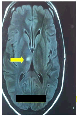
Figure 2: Magnetic resonance imaging of the brain
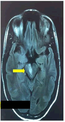
Figure 3: Axial T2-weighted MRI showing a pontine infarct (yellow arrow)
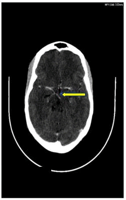
Figure 4: Contrasted CT image. Showing ‘beading’ of the paramedian blood vessels supplying the midbrain (signs of vasculitis)
The axial view shows the right hyperdense lesion at the paramedian thalami caused by an ischemic infarct secondary to HIV vasculitis.
The axial view shows a hyperdense lesion on the right midbrain caused by ischemic stroke due to HIV vasculitis.
The cardiology team requested a cardiac review. A cardiac ultrasound (done the day after admission) showed an ejection fraction of 75%. No signs of valvulopathy or effusion were present.
Because our patient was not a candidate for thrombolytics because his symptoms began twenty-two hours prior to first medical contact, which falls outside of the 4.5-hour therapeutic window for intravenous thrombolysis using tissue plasminogen. Her negative CTA precluded his candidacy for endovascular therapy; pharmacological management included antiretroviral therapy, antiplatelet aggregation, symptomatic and supportive therapy. Therefore, drug therapy was administered as follows: Vitamin B12 supplementation (1000 μg IM daily for five days in the first week, then weekly for five weeks, aspirin (150 mg daily), enoxaparin (40 mg s/c daily), simvastatin (20 mg daily), pyridoxine (50 mg daily), thiamine (100 mg daily). The patient continued the chronic medication (hydrochlorothiazide 12.5 mg, enalapril 5 mg and TDF/ FTC/EFV 300/200/600 mg daily). Physiotherapy and occupational therapy are actively working with the patient (TJ4). The patient received rehabilitation in our ward for two weeks. The right-sided hemiataxia improved, but rightsided power remained 4/5. She was referred to her base hospital to continue rehabilitation and a follow-up date with us in one month’s time.
Discussion
Here we report a case of a 41-year-old woman with rightsided TMS. Unilateral TMS is uncommon; its incidence remains unknown, but one study reported that it accounts for about 0.6-1% of midbrain ischaemic strokes and is often accompanied by other posterior circulation infarcts [32].
Baran et al., conducted an observational study in 2018, which showed a male predominance. The study also showed that, from an etiological point of view, the most common cause was extensive atherosclerosis, followed by cardioembolism, with small-vessel disease being the least common [33]. The main risk factors associated with extensive atherosclerosis are hypertension, diabetes, hyperlipidaemias, smoking, and previous history of stroke. The main risk factors for cardio-embolism in these patients are atrial fibrillation [33]. Patients who are suffering from peripheral vascular disease and coronary artery disease are at risk [34].
HIV is a risk factor for stroke [35] and is associated with advanced disease [36]. Numerous mechanisms have been proposed to explain this. A systematic review done by Addallah et al., reported that this could be due to HIV-associated opportunistic infections, HIV-induced coagulopathy, and chronic inflammatory processes that can accelerate atherosclerosis [37]. Another systematic review by Bogorodskaya et al., also reported that some antiretrovirals (lopinavir, indinavir, and abacavir) were also associated with an increased risk of stroke [38].
Lesions of the midbrain can present as distinct syndromes. However, because of the structures’ close organisation, there can be considerable overlap of these syndromes. The neurological manifestation will depend on which area of the midbrain is affected, whether one half or both halves are involved, and whether adjacent structures (thalamus, pons, cerebellum) are also involved. The symptoms may include, but are not limited to, low equilibrium, weakness on one or both sides of the body, diplopia, and slurred speech [39]. The most common examination findings include ataxia, limb weakness, dysarthria, sensory disturbance, oculomotor findings (3rd nerve palsy, internuclear ophthalmoplegia), and dysarthria [40]. The exact pattern will depend on the area involved and whether surrounding structures are also involved (thalamus, pons, medulla, etc.). There are midbrain syndromes, which include, among others, Weber syndrome, Claude’s syndrome, Nothnagel syndrome, and Benedikt’s syndrome. Benedikt’s syndrome presents with a contralateral rubral tremor, which she does not have.
Weber syndrome is a result of a lesion involving the ventromedial area of the midbrain. They present with ipsilateral 3rd nerve palsy with contralateral hemiplegia. Our patient has Claude’s syndrome, which presents with ipsilateral 3rd nerve palsy and contralateral cerebellar ataxia due to a dorsal tegmental lesion that involves the 3rd nerve nucleus/fibres and either the red nucleus, superior cerebellar peduncle, or brachium conjunctivum [37]. Benedikt’s syndrome is due to a lesion involving the tegmentum. It presents with ipsilateral 3rd nerve palsy and contralateral ataxia, but there is also involvement of the fibres of the corticospinal tract, which will result in contralateral hemiparesis [41]. When assessing these kinds of patients, it is essential to ascertain a good history and physical examination and check the National Institutes of Health Stroke scale [42]. Imaging to confirm the diagnosis is mandatory. CT or MRI angiography is usually requested to identify stenosed vessels or other possible vascular problems. Blood workup for stroke is compulsory and includes, but is not limited to, full blood count, renal function tests, international normalised ratio, lipid profile, HIV-ELISA, and, if young, a thrombophilia screen, Antinuclear antibodies, and glycosylated haemoglobin. ECG to rule out possible atrial fibrillation and transthoracic or even transoesophageal echocardiography to identify cardiac causes.
The management approach depends on the aetiology of the stroke. If the infarct is ischaemic, the reviewed literature recommends thrombolysis for posterior circulation strokes that meet the established criteria [43]. The benefits of mechanical thrombectomy are not yet well established, but it can be performed [44]. Then, after the acute period, it is crucial to manage the risk factors and causes. Then treat the risk factors such as arterial hypertension, diabetes mellitus, hyperlipidaemia, and secondary prophylaxis. If there is a cardiac cause, then it should be treated. A multidisciplinary approach is vital for patients presenting with TMS. The stroke team should include dietitians, physiotherapists, speech therapists, occupational therapists, and social workers, in addition to the medical specialists.
Risk factors for developing stroke in our patient were hypertension, HIV, and hyperlipidaemia. We had done an extensive workup to rule out other possible contributors to a stroke. The patient also had contralateral hemiparesis and hemiataxia with bilateral Babinski and hyperreflexia on both lower limbs. Her blood workup showed that she was virally suppressed and had hyperlipidaemia. The MRI and CT angiograms showed evidence of an infarct involving the ventromedial midbrain and thalamus, with no other lesions. Cardiovascular investigations ruled out a cardiac source of the infarct. In this patient, hypertension, HIV infection, and hyperlipidaemia predisposed her to the stroke. The patient is markedly younger than one would typically expect for a TMS (median age around 64 years) [32]. Of note in our patient is the presence of a bilateral Babinski sign, which is never seen in patients with Claude’s syndrome.
Generally, strokes involving the posterior circulation have a higher mortality rate than those involving the anterior circulation, unless they involve the smaller blood vessels [41], as in our case. The present case is unique, among other reasons, owing to the bilateral Babinski sign and the absence of thalamic manifestations, in the absence of other lesions affecting different segments of the brainstem and the spinal cord. The patient’s age (41 years) also makes this case uncommon. The patient’s leading risk factor is HIV vasculitis, which has not been implicated for TMS from the literature reviewed. In our setting, the commonest causes of infectious vasculitis are HIV/AIDS, tuberculosis, neurocysticercosis, and neurosyphilis, which is quite different from other countries.
Therefore, HIV vasculitis should be the first differential diagnosis in our region. However, we could not confirm this aetiology; we decided to use the terminology: “an inpatient with HIV” because it was confirmed. We did not find signs of Middle Longitudinal Fascicle (MLF) typical syndrome. MLF syndrome, secondary to ischemic stroke affecting only the mesencephalon, is a rare occurrence [45,46]. We were unable to identify the cause of the bilateral Babinski signs in this case. This patient did not present thalamic characteristics despite the ischemic lesion in the right thalamus, despite the midbrain’s role over the thalamus. The modulation of thalamic neurons is necessary to control adaptive behaviour mediated by midbrain cholinergic transmission. The central cholinergic neurons in the mesencephalon are in the pedunculopontine nucleus and the laterodorsal tegmental nucleus, which provide dense innervation of the thalamus. Recently, Huerta-Ocampo et al., confirmed that midbrain cholinergic neurons could innervate all thalamic nuclei. They also found that these axons are topographically well organised and provide a segregated innervation of the thalamic nuclei [47]. Therefore, we expected some evidence of thalamic dysfunction in this patient. However, these midbrain ischemic lesions did not cause abnormal behaviour or thalamic manifestations in our patient, a novel finding.
Brief comments on the thalamus
Blood supply to the thalamic nucleus includes several branches of the Posterior Cerebral Artery (PCA) which are shown in Figure 5.
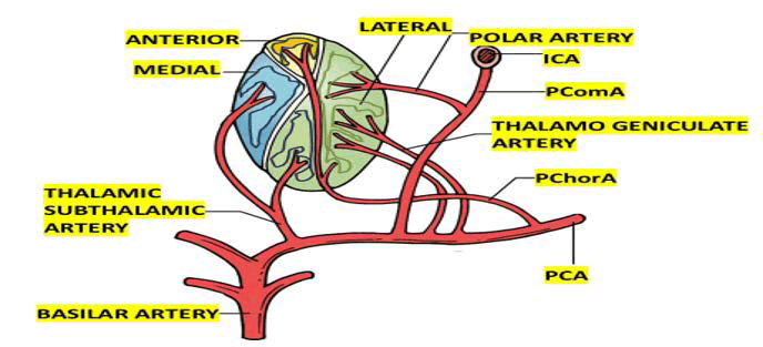
Figure 5: Shows all the branches supplying the anterior, medial and lateral aspect of the thalamus
The thalamus is a diencephalic nuclear complex that plays a key role in memory, emotions, regulation of the sleep wake cycle, executive processes, cortical alertness, and sensorimotor integration [48]. The lateral, medial, and posterior thalamic regions are primarily supplied by the vertebrobasilar system through branches of the posterior cerebral and posterior communicating arteries [49].
Our vascular configuration is a new variant, as the thalami are usually supplied by multiple perforating arteries. The graphical representation of the AOP is shown in Figure 6.
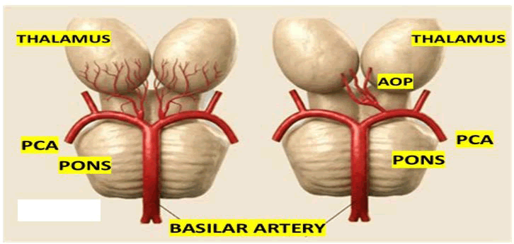
Figure 6: Shows the commonest presentation of AOP supplying both thalamic regions
The presence of occlusion of the AOP explains the clinical manifestation related to bilateral thalamic injury, often with devastating neurological outcomes. However, in our case the HIV vasculitis may affect only some branches and keep other impaired which explain the asymmetrical reduced blood supply to the bilateral thalamus and the presence of bilateral upper motor neuron signs without lack of other thalamic features such as disturbances of consciousness, memory, motor coordination, aphonia, apathy, failure to comprehend, severe hypersomnolence and akinetic mutism supported by bilateral thalamic and midbrain involvement as has been proposed by other authors [50].
The pathophysiology of AOP infarction primarily involves aetiologies like atherosclerosis, small vessel disease, and embolic events secondary to cardioembolism among other sources. However, HIV-infectious vasculitis as a cause has not been reported to our knowledge.
There are four types of IS affecting the Thalamicmesencephalic region as listed below:
• Bilateral paramedian thalamic and rostral midbrain infarction. The main cause of bilateral paramedian thalamic infarctions was small artery disease (60%), followed by cardioembolism (40%). A well-defined clinical picture is shown in bilateral paramedian thalamic artery infarcts. These patients had disorders consisting of co altered mental status, memory dysfunctions, and various types of vertical gaze paresis and psychological changes.
• Bilateral paramedian thalamic infarction without midbrain involvement. This variant occurs in about 38% of cases. The lesion directly impacts the paramedian thalami, yet the development of midbrain arteries remains unchanged. The variant may be observed in changes of mood, cognitive, neurological, and cognitive-shift forms. This is because the important motor tracts are preserved in the remaining midbrain, and thus the prognosis is slightly better than in cases where both the thalami and the midbrain are impaired.
• Bilateral paramedian and anterior thalamic infarction with midbrain involvement. Li et al., reported that this variant is present in 14% of all patients. This affects all the midbrain, paramedian, and anterior thalamic regions. Its features may include prolonged loss of consciousness, memory impairment, changes in behaviour, and visual acuity [51].
• Bilateral paramedian and anterior thalamic infarction without midbrain involvement. This variant is seen in roughly 5% of cases, making it the least common form. It spreads across the anterior and paramedian subsections and does not involve the midbrain [51]. It may present as changes in memory, sensory examination, and cognitive function, which are universally impaired except in areas governed by midbrain oculomotor and motor functions.
The topographic lesion at the level of bilateral thalami and rostral midbrain are represented in Figure 7.
Four neurovascular anatomical variants have been described for the thalamus and midbrain (Table 2) [52].
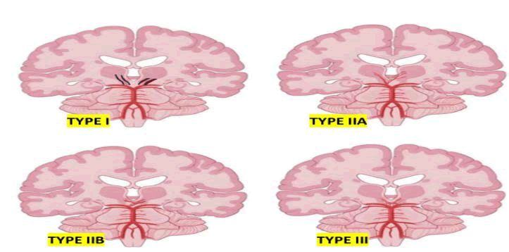
Figure 7: This illustration demonstrates four types of AOP infarction. (A) It shows paramedian arteries stemming from each PCA (Type I). (B) It shows paramedian arteries arising from a communicating branch between the PCAs (Type IIa). (C) It shows the AOP arises unilaterally from a PCA and gives rise to the paramedian arteries (Type IIb). (D) It shows paramedian arteries arising from a communicating branch between the PCAs (Type III)
| Type | Affected artery | Frequency | Arising segment | Point E |
| Type I | Two paramedian arteries | Common | Proximal P1 | EACH PCA |
| Type IIa | Both paramedian | Rare | Proximal P1 of left/right | PCA |
| Type IIb | Single trunk (AOP) | Commonest | Unilateral from P1 | Bifurcating to supply paramedian thalamus and bilateral rostral midbrain |
| Type III | Communicating artery | Less common | Proximal aspect (P1) of both left and right PCA | From paramedian arteries originate |
Table 2: Variants of the paramedian thalamic arterial supply, including the Artery of Percheron (AOP), with their frequency, site of origin, and vascular territories supplied
The commonest variant of Percheron is type IIb, which supplies the paramedian thalami and the rostral midbrain [53]. Other variants of AOP are shown in Figure 8.
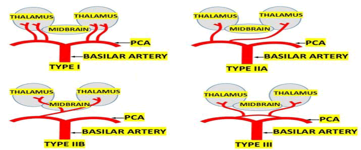
Figure 8: Schematic illustration of the four anatomical variants of paramedian thalamic artery supply, including the artery of Percheron (Type IIb), arising from the posterior cerebral artery and supplying the bilateral thalami and rostral midbrain
Type IIb occlusion may cause a bilateral paramedian thalamic stroke, while the rostral midbrain and anterior thalamus are involved in 57% and 19% cases, respectively. However, the less common type III variant prevented the bilateral extension of infarction and involved the territory of tuberothalamic and paramedian perforating arteries [54- 56].
Most patients with an AOP IS have progressive drowsiness, progressing to a coma, and these clinical presentations may vary due to the disparate functions of the implicated structures [57].
In cases presenting with midbrain involvement, vertical gaze palsy and ataxia can be observed, along with behavioural and memory deficits related to dysfunction of the hippocampal region or its connections, motor dysfunction, and speech difficulties [57-59].
We represent the thalamic-mesencephalic lesion in Figure 9.
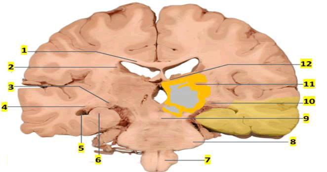
Figure 9: Shows an extensive ischemic lesion in the thalamic region. 1) Body of corpus callosum, 2) Body of caudate nucleus, 3) Lateral geniculate nucleus of the thalamus, 4) Tail of caudate nucleus, 5) Inferior horn of the lateral ventricle, 6) Hyppocampus, 7) Medulla, 8) Middle cerebellar peduncle, 9) Decussation of the superior cerebellar peduncle, 10) Medial geniculate body of the thalamus, 11) Pulvinar thalamus, 12) Crus of fornix
In our case unilateral thalamic lesion was confirmed by imaging and we hypothesised that the contralateral thalamus was also damage which explain bilateral Babinski sign and alterated level of consciousness due to involvement the diencephalic structures of the activating reticular formation system. Therefore, as we before mentioned the presence of bilateral Babinski signs is due to involvement of upper motor neuron fibres close to the thalami bilaterally, with a lack of complete thalamic manifestation reflecting partial impairment of the left thalamus, attributable to the absence of damage caused by HIV vasculitis on the less unaffected side. It is important to highlight that the thalamic branches are composed of a group of perforating arteries of the thalamus that originate from the posterior communicating and cerebral arteries, including the thalamogeniculate and medial and lateral posterior choroidal arteries, which are not affected by HIV in a symmetrical and generalised pattern. On the other hand, there are several variants of blood supply to the thalamus, and the identification of the potential presence and infarction of an AOP is important in early diagnosis and drug therapy of IS affecting the thalami and midbrain, especially because of the unusual and variable presentation of these forms of ischemic IS [60,61].
AOP stroke following surgical clipping of anterior circulation aneurysms has not been documented in the literature [62].
In variant I, the most common variant, symmetrical perforating arteries arise from both left and right PCAs. Variant IIa is a rare asymmetrical configuration in which perforating arteries originate exclusively from the P1 segment of one PCA. Variant IIb, which is also uncommon, is the classic AOP, which arises as a single arterial trunk from the P1 segment of one PCA and bifurcates to supply the bilateral paramedian thalami and the rostral midbrain; occlusion of this variant produces a characteristic bilateral thalamic infarction pattern. In variant III (an arcade variant), multiple small perforating branches emerge from an arterial arch connecting the P1 and P2 segments of the PCA. Classically, AOP infarct is quite often characterised by the clinical trial of altered mental status, vertical gaze palsy, and memory impairment, though presentation can vary widely, and symptoms such as hypersomnolence, akinetic mutism, and oculomotor dysfunction may also occur [63].
These non-specific manifestations and a variety of manifestations very often lead to misdiagnosis or delayed clinical suspicion, particularly in younger patients without traditional vascular risk factors. These several types of presentation are attributable to the central role of the thalamus in consciousness, cognition, and motor integration, and it underscores the importance of clinical vigilance and advanced imaging for recognising this rare yet critical subtype of stroke.
We hypothesised that the clinical manifestations change depending on the thalamic subregions involved, and that they may overlap with midbrain syndromes, which are more pronounced in cases of vasculitis presenting segmental involvement along the vessel wall over its length, thereby leading to a wide variety of clinical manifestations according to the affected vascular territory.
Brief comments on differential diagnosis of AOP infarction
To distinguish AOP infarction from other causes of bilateral thalamic lesions, it is important to consider the overlapping imaging and clinical features in conditions like Wernicke’s encephalopathy, viral encephalitis, cerebral sinus thrombosis, and metabolic disorders (Table 3) [64,65].
This table was modified from [66].
| Category | Condition | Clinical features | Imaging findings |
| Vascular | Artery of Percheron infarct | Somnolence, memory loss, vertical gaze palsy | Bilateral paramedian thalamic ± midbrain infarcts on DWI/FLAIR |
| Cerebral venous sinus thrombosis (e.g., vein of Galen) | Headache, altered consciousness, seizures | Bilateral thalamic edema, hyperintense veins, venous infarction pattern on MRV | |
| Top of the basilar syndrome | Coma, quadriplegia, oculomotor palsy | Infarcts in the thalami, midbrain, and occipital lobes | |
| Neoplastic | Bilateral thalamic glioma | Movement disorders, cognitive decline | Diffuse T2 hyperintensity, mass effect without contrast enhancement |
| Infectious | Japanese encephalitis | Fever, confusion, seizures, focal deficits | Bilateral thalamic hyperintensities with oedema |
| West Nile virus encephalitis | Flu-like symptoms, confusion, tremor | T2 hyperintensity in the thalami, basal ganglia, and midbrain | |
| Toxoplasmosis (immunocompromised) | Fever, focal neurologic signs | Ring-enhancing lesions in deep gray matter (thalamus, basal ganglia) | |
| Metabolic/Toxic | Wernicke’s encephalopathy | Ataxia, ophthalmoplegia, confusion | Symmetric thalamic hyperintensity on T2/FLAIR; affects mammillary bodies, periaqueductal gray |
| Osmotic demyelination (extrapontine) | Altered mental status after rapid Na+ correction | Bilateral T2 hyperintensities in the thalami and basal ganglia | |
| Wilson’s disease | Movement disorders, psychiatric symptoms | T2 hyperintensity in the thalami and basal ganglia; "face of giant panda" sign | |
| Leigh syndrome (mitochondrial) | Childhood onset, psychomotor regression, respiratory failure | T2 hyperintensity in the thalami, brainstem, and basal ganglia | |
| Gangliosidosis | Developmental delay, regression, spasticity | Symmetric white matter changes, thalamic calcification | |
| Autoimmune/other | Posterior reversible encephalopathy | Seizures, visual disturbance, hypertension | Vasogenic edema in occipital and parietal lobes ± thalami |
| Creutzfeldt-Jakob disease | Rapid dementia, myoclonus | Pulvinar sign; high DWI signal in the thalami and basal ganglia | |
| Fatal familial insomnia | Progressive insomnia, autonomic failure | Thalamic hypometabolism on PET; subtle thalamic atrophy | |
| Note: DWI: Diffusion-Weighted Imaging; FLAIR: Fluid-Attenuated Inversion Recovery; MRV: Magnetic Resonance Venography; PET: Positron Emission Tomography | |||
Table 3: Differential diagnosis of bilateral thalamic lesions
Brief comments on confirmation and management
Successful management of AOP stroke depends on accurate, confident early identification of AOP infarction, which requires a high index of clinical suspicion and advanced imaging techniques. In most cases, Computed Tomography (CT) is normal in the acute phase, whereas modalities such as MRI with Diffusion-Weighted Imaging (DWI) and Fluid-Attenuated Inversion Recovery (FLAIR) sequences are more sensitive to the subtle bilateral paramedian thalamic changes typical of this condition [67].
The AOP is frequently invisible on conventional MRA or Computed Tomography Angiography (CTA) due to its deep-seated location and small calibre, thereby posing a significant diagnostic challenge. In our case, DWI and FLAIR sequences confirmed bilateral thalamic infarction with a visible AOP on MRA, which is relatively uncommon given the low angiographic sensitivity reported in the literature [68].
In our patient the CT angiographic study confirmed the signs of vasculitis only in one side of the branches of the AOP as we represented in Figure 10.
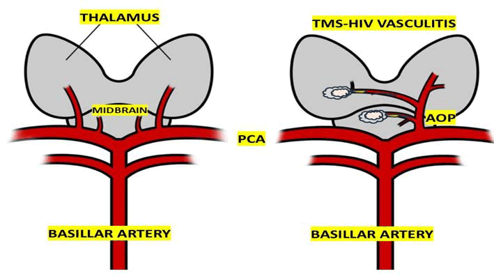
Figure 10: Represent our hypothesis on the affected vascular territory of the AOP causes by HIV-vasculitis involving the thalamic branches unilaterally
In cases presenting within the therapeutic window, thrombolysis has been reported to improve outcomes [68]. However, delayed recognition due to the often non-focal and non-lateralizing nature of symptoms frequently leads to missed opportunities for reperfusion therapies.
The management of AOP occlusion typically includes antiplatelet therapy and lipid-lowering agents, in addition to management of detected paroxysmal atrial fibrillation and transthoracic echocardiography with a bubble study to assess for cardiac sources of embolism, a young stroke blood panel to rule out antiphospholipid syndrome, JAK2 mutation, or paroxysmal nocturnal haemoglobinuria.
Brief comments on thrombolytic therapy
Recently, some authors reported a 70-year-old male comatose patient who presented within the thrombolysis window. CT head and CT angiogram were normal, and he was successfully thrombolysed. Urgent MRI with DWI was arranged, demonstrating bilateral paramedian thalamic diffusion restriction, reported as Percheron artery infarction [69].
Conclusion
To our knowledge, this is the first patient presenting with unilateral TMS HIV vasculitis-related due to partial occlusion of AOP branches with bilateral Babinski signs, and without complete thalamic manifestations reported in the medical literature. In young patients presenting with unilateral TMS, HIV vasculitis is one of the etiological diagnoses to be considered. Still, we recommend an extensive investigation based on a series of cases to support this postulate.
As far as we know, this is the first study that graphically represent the elements involved in the pathogenesis of midbrain stroke secondary to HIV vasculitis and its mechanism of reperfusion at the rostral midbrain.
Acknowledgment
To thanks to Dr. Sibi Joseph for his collaboration on the management of our case series.
Ethics Statement
The current review does not qualify for ethical approval.
Patient Privacy
All information related to identification of patients were removed to provide complete anonymity.
Conflicts of Interest
Authors report no conflicts of interest
References
- M. Kashif, A. Hamed, I. Bozkurt, N. Montemurro, B. Chaurasia, et al. The clinical significance of artery of Percheron in cerebral ischemia: A summary overview, Ann Med Surg (Lond), 87(2025):4310–4315.
[Crossref] [Google Scholar] [PubMed]
- O.A.M. Kheiralla, Artery of Percheron infarction a rare anatomical variant and a diagnostic challenge: Case report, Radiol Case Rep, 16(2021):22–29.
[Crossref] [Google Scholar] [PubMed]
- A. Sandvig, S. Lundberg, J. Neuwirth, Artery of Percheron infarction: A case report, J Med Case Rep, 11(2017):221.
[Crossref] [Google Scholar] [PubMed]
- O. Atallah, Y.F. Almealawy, A.S. Alabide, Navigating the clinical landscape of artery of Percheron infarction: A systematic review, Eneurol Sci, 37(2024):100521.
[Crossref] [Google Scholar] [PubMed]
- J.C. Arcidiacono, A. Whitsett, K. Callagy, R. Colella, The unlucky variant: Artery of Percheron infarction, Cureus, 17(2025):e84582.
[Crossref] [Google Scholar] [PubMed]
- V. Nirmalan, Artery of Percheron syndrome from a possible paradoxical embolus in the intensive care unit: an easily missed diagnosis in intubated patients, Cureus, 18(2026):e109077.
[Crossref] [Google Scholar] [PubMed]
- S. Ansar, R. Umer, C.M.A.A. Gamage, M. Sharjeel, Z. Ammer, et al. Artery of Percheron infarction presenting as altered consciousness in an elderly patient: A case report, Cureus, 17(2025):e87588.
[Crossref] [Google Scholar] [PubMed]
- N. Zuñiga Rivera, C.R. Vazquez Sotelo, A. Reyes Valdes, A. Albarrán Sánchez, Bilateral thalamic infarction due to artery of Percheron occlusion in a young patient with hyperhomocysteinemia and vertebral artery dissection, Cureus, 17(2025):e92282.
- N.R. Barefoot, A.R. Cunningham, H.E. Behm, A.W. Ju, M.S. Peach, et al. Acute artery of Percheron infarction: A case report highlighting diagnostic challenges and management, Cureus, 17(2025):e82072.
[Crossref] [Google Scholar] [PubMed]
- Y. Matsushima, T. Sanada, A. Tucker, M. Kinoshita, T. Kimura, et al. Mechanical thrombectomy for top of basilar artery occlusion and delayed distal thrombus migration involving the artery of Percheron: Illustrative case, J Neurosurg Case Lessons, 9(2025):CASE25171.
[Crossref] [Google Scholar] [PubMed]
- M. NavickaitÃÂ, A. Vilionskis, A. DapkutÃÂ, K. RyliškienÃÂ, Artery of Percheron stroke: A case report, Acta Med Litu, 32(2025):145–152.
[Crossref] [Google Scholar] [PubMed]
- A.Z. Thomas, A. Krishnan, H. Joseph, A. Sreekumar, V. Jayachandran, et al. Artery of Percheron infarction: Diagnostic and clinical insights from two cases, Cureus, 17(2025):e95907.
[Crossref] [Google Scholar] [PubMed]
- W.A. Sanfelippo, M. Oley, H. Harrelson, N. Vilar, Artery of Percheron infarct with multiple cranial nerve palsies and Horner syndrome, Am J Ophthalmol Case Rep, 40(2025):102458.
[Crossref] [Google Scholar] [PubMed]
- M.T.H. Zon, K.W.Y. Lwin, O. Owolabi, S. Omer, The unusual suspect: Artery of Percheron occlusion in a young healthy patient, Cureus, 17(2025):e96953.
[Crossref] [Google Scholar] [PubMed]
- X.F. Nong, X. Cao, X.L. Tan, L.Y. Jing, H. Liu, et al. Percheron syndrome with memory impairment as chief manifestation: A case report, World J Clin Cases, 13(2025):98937.
[Crossref] [Google Scholar] [PubMed]
- J. Nagasawa, M. Hozumi, T. Yokoyama, M. Ogawa, J. Ebina, et al. Posterior communicating artery configuration and laterality of thalamic and lenticulostriate infarction, Neurol Int, 18(2026):61.
[Crossref] [Google Scholar] [PubMed]
- S. Tian, P.A. Patel, D.V. Giurgiutiu, Artery of Percheron stroke causing sudden severe decreased consciousness, Stroke Vasc Interv Neurol, 5(Suppl 1)(2025):e70000_202.
- M.D. Hussen, Z. Tabassum, Artery of Percheron infarction and a very rare finding of ping pong gaze: A case report, Radiol Case Rep, 21(2026):1658–1662.
- S. Marinkovic, M. Milisavljevic, M. Kovacevic, Interpeduncular perforating branches of the posterior cerebral artery: Microsurgical anatomy of their extracerebral and intracerebral segments, Surg Neurol, 26(1986):349–359.
[Crossref] [Google Scholar] [PubMed]
- A. Pedroza, M. Dujovny, J.I. Ausman, Microvascular anatomy of the interpeduncular fossa, J Neurosurg, 64(1986):484–493.
[Crossref] [Google Scholar] [PubMed]
- P.E.J. Caballero, Bilateral paramedian thalamic artery infarcts: Report of 10 cases, J Stroke Cerebrovasc Dis, 19(2010):283–289.
[Crossref] [Google Scholar] [PubMed]
- N.A. Lazzaro, B. Wright, M. Castillo, Artery of Percheron infarction: Imaging patterns and clinical spectrum, AJNR Am J Neuroradiol, 31(2010):1283–1289.
[Crossref] [Google Scholar] [PubMed]
- Y.M. Song, Topographic patterns of thalamic infarcts in association with stroke syndromes and aetiologies, J Neurol Neurosurg Psychiatry, 82(2011):1083–1086.
[Crossref] [Google Scholar] [PubMed]
- A. Arauz, H.M. Patiño-Rodríguez, J.C. Vargas-González, Clinical spectrum of artery of Percheron infarct: Clinical-radiological correlations, J Stroke Cerebrovasc Dis, 23(2014):1083–1088.
[Crossref] [Google Scholar] [PubMed]
- A. Förster, I. Nölte, H. Wenz, Anatomical variations in the posterior part of the circle of Willis and vascular pathology in bilateral thalamic infarction, J Neuroimaging, 24(2014):325–330.
[Crossref] [Google Scholar] [PubMed]
- Z. Xu, L. Sun, Y. Duan, Assessment of Percheron infarction in images and clinical findings, J Neurol Sci, 383(2017):87–92.
[Crossref] [Google Scholar] [PubMed]
- B.L. Man, Y.P. Fu, See-saw nystagmus, convergence-retraction nystagmus, and contraversive ocular tilt reaction from a paramedian thalamomesencephalic infarct, BMJ Case Rep, 2014(2014):bcr2014206851.
[Crossref] [Google Scholar] [PubMed]
- M. Versino, F. Simonetti, M.G. Egitto, Lateral gaze synkinesis on downward saccade attempts with paramedian thalamic and midbrain infarct, J Neurol Neurosurg Psychiatry, 67(1999):696–697.
[Crossref] [Google Scholar] [PubMed]
- P. Homeyer, P. Cornu, L. Lacomblez, A special form of cerebral lacunae: Expanding lacunae, J Neurol Neurosurg Psychiatry, 61(1996):200–202.
[Crossref] [Google Scholar] [PubMed]
- L.A. Benjamin, A. Bryer, H.C.A. Emsley, HIV infection and stroke: Current perspectives and future directions, Lancet Neurol, 11(2012):878–890.
[Crossref] [Google Scholar] [PubMed]
- L. Kouassi, M. Doumbia-Ouattara. Acute onset vascular dementia with bi-thalamic infarct in an HIV-positive subject, Am J Case Rep, 18(2017):1145–1147.
[Crossref] [Google Scholar] [PubMed]
- J. Bogousslavsky, F. Regli, P. Maeder, The etiology of posterior circulation infarcts: A prospective study using magnetic resonance imaging and magnetic resonance angiography, Neurology, 43(1993):1528–1533.
[Crossref] [Google Scholar] [PubMed]
- G. Baran, T.O. Gultekin, O. Baran, Association between etiology and lesion site in ischemic brainstem infarcts: A retrospective observational study, Neuropsychiatr Dis Treat, 14(2018):757–766.
[Crossref]
- A.C.L. Neto, R. Bittar, G.S. Gattas, Pathophysiology and diagnosis of vertebrobasilar insufficiency: A review of the literature, Int Arch Otorhinolaryngol, 21(2017):302–307.
[Crossref] [Google Scholar] [PubMed]
- L. Benjamin, S. Khoo, HIV infection and stroke, Handb Clin Neurol, 152(2018):187–200.
- F. Chow, P. Bacchetti, A. Kim, Effect of CD4+ cell count and viral suppression on the risk of ischemic stroke in HIV infection, AIDS, 28(2014):2573–2577.
[Crossref] [Google Scholar] [PubMed]
- A. Abdallah, J.L. Chang, C.B. O'Carroll, Stroke in HIV-infected individuals in sub-Saharan Africa (SSA): A systematic review, J Stroke Cerebrovasc Dis, 27(2018):1828–1836.
- M. Bogorodskaya, F.C. Chow, V.A. Triant, Stroke in HIV, Can J Cardiol, 35(2019):280–287.
- L. Pirau, F. Lui, J.P. Mohr, L.R. Caplan, Vertebrobasilar insufficiency. In: Stroke: Pathophysiology, Diagnosis, and Management, Philadelphia: Elsevier Saunders, (2011):446–484.
- J.S. Kim, J. Kim, Pure midbrain infarction: Clinical, radiologic and pathophysiologic findings, Neurology, 64(2005):1227–1232.
[Crossref] [Google Scholar] [PubMed]
- K. Ruchalski, G.M. Hathout, A medley of midbrain maladies: A brief review of midbrain anatomy and syndromology for radiologists, Radiol Res Pract, 2012(2012):258524.
[Google Scholar] [PubMed]
- L. KhimKwah, J. Diong, National Institutes of Health Stroke Scale (NIHSS), J Physiother, 60(2014):61.
[Crossref] [Google Scholar] [PubMed]
- J.O. Filho, O.B. Samuels, Approach to reperfusion therapy for acute ischemic stroke, (2020).
- W.J. Powers, A.A. Rabinstein, T. Ackerson, Guidelines for the early management of patients with acute ischemic stroke: 2019 update to 2018: A guideline for healthcare professionals from the American Heart Association/American Stroke Association, Stroke, 50(2019):e344–e418.
- S.S. Yeo, S.H. Jang, J.W. Kwon, Three-dimensional identification of the medial longitudinal fasciculus in the human brain: A diffusion tensor imaging study, J Clin Med, 9(2020):1340.
[Crossref] [Google Scholar] [PubMed]
- P.S. Kochar, Y. Kumar, P. Sharma, Isolated medial longitudinal fasciculus syndrome: Review of imaging, anatomy, pathophysiology and differential diagnosis, Neuroradiol J, 31(2018):95–99.
[Crossref] [Google Scholar] [PubMed]
- I. Huerta-Ocampo, H. Hacioglu-Bay, D. Dautan, Distribution of midbrain cholinergic axons in the thalamus, eNeuro, 7(2020):ENEURO.0454-19.2019.
[Crossref] [Google Scholar] [PubMed]
- E. Carrera, J. Bogousslavsky, The thalamus and behavior: Effects of anatomically distinct strokes, Neurology, 66(2006):1817–1823.
[Crossref] [Google Scholar] [PubMed]
- S. Bordes, C. Werner, M. Mathkour, Arterial supply of the thalamus: A comprehensive review, World Neurosurg, 137(2020):310–318.
[Crossref] [Google Scholar] [PubMed]
- A. Ciacciarelli, I. Francalanza, F. Giammello, Prevalence, clinical features, and radiological pattern of artery of Percheron infarction: A challenging diagnosis, Neurol Sci, 44(2023):2413–2419.
[Crossref] [Google Scholar] [PubMed]
- X. Li, N. Agarwal, D.R. Hansberry, Contemporary therapeutic strategies for occlusion of the artery of Percheron: A review of the literature, J Neurointerv Surg, 7(2015):95–98.
[Crossref] [Google Scholar] [PubMed]
- N.A. Lazzaro, B. Wright, M. Castillo, Artery of Percheron infarction: Imaging patterns and clinical spectrum, AJNR Am J Neuroradiol, 31(2010):1283–1289.
[Crossref] [Google Scholar] [PubMed]
- M. Zedde, I. Grisendi, F. Assenza, Type III Percheron’s variant in thalamic-mesencephalic infarction: The unexpected anastomosis, Surg Radiol Anat, 46(2024):1769–1774.
[Crossref] [Google Scholar] [PubMed]
- M. Zedde, I. Grisendi, F. Assenza, Type III Percheron’s variant in thalamic-mesencephalic infarction: The unexpected anastomosis, Surg Radiol Anat, 46(2024):1769–1774.
[Crossref] [Google Scholar] [PubMed]
- P. Zanzmera, K. Jain, S. Garg, Successful thrombolysis in artery of Percheron infarction: Grabbed an extremely rare opportunity, Neurol India, 72(2024):632–634.
[Crossref] [Google Scholar] [PubMed]
- J.C. Pérez-Cruz, M.A. Macías-Duvignau, G. Reyes-Soto, Latex vascular injection as method for enhanced neurosurgical training and skills, Front Surg, 11(2024):1366190.
[Crossref] [Google Scholar] [PubMed]
- N. Jaafar, R. Sharma, N. Parkash, Decoding altered consciousness: An artery of Percheron stroke, Cureus, 16(2024).
[Crossref] [Google Scholar] [PubMed]
- B. Zhang, X. Wang, C. Gang, Acute Percheron infarction: A precision learning, BMC Neurol, 22(2022):207.
[Crossref] [Google Scholar] [PubMed]
- N. Phate, T. Pawar, A. Andhale, E.P. Nolley, N. Parkash, et al. Artery of Percheron stroke: A case report with a diagnostic challenge, Cureus, 14(2022).
[Crossref] [Google Scholar] [PubMed]
- A. Kichloo, S.M. Jamal, E.A. Zain, Artery of Percheron infarction: A short review, J Investig Med High Impact Case Rep, 7(2019):2324709619867355.
[Crossref] [Google Scholar] [PubMed]
- G. Reyes-Soto, J.C. Pérez-Cruz, L. Delgado-Reyes, The vertebrobasilar trunk and its anatomical variants: A microsurgical anatomical study. Diagnostics (Basel) 14(2024):534.
[Crossref] [Google Scholar] [PubMed]
- M. Chen, X. Xia, L. Chen, L. Yang, Z. Li, et al. Case report: Artery of Percheron infarction following surgical clipping of multiple intracranial aneurysms. Front Surg. 2025(12):1623891.
[Crossref] [Google Scholar] [PubMed]
- M.A. Garcia-Grimshaw, M. Peschard-Franco, F.A. Gutierrez-Manjarrez, Bilateral thalamic ischemic stroke secondary to occlusion of the artery of Percheron, Cureus, 10(2018):e2676.
- H.B. Bhattarai, S.R. Dahal, M, Uprety, Bilateral thalamic infarct involving artery of Percheron: A case report, Ann Med Surg (Lond), 85(2023):4613–4618.
[Crossref] [Google Scholar] [PubMed]
- A.B. Smith, J.G. Smirniotopoulos, E.J. Rushing, S.J. Goldstein, Bilateral thalamic lesions, AJR Am J Roentgenol, 192(2009):W53–W62.
[Crossref] [Google Scholar] [PubMed]
- M.T.H. Zon, K.W.Y. Lwin, O. Owolabi, S. Omer, The unusual suspect: Artery of Percheron occlusion in a young healthy patient, Cureus, 17(2025):e96953.
[Crossref] [Google Scholar] [PubMed]
- O. Atallah, Y.F. Almealawy, A.S. Alabide, Navigating the clinical landscape of artery of Percheron infarction: A systematic review, eNeurologicalSci, 37(2024):100521.
[Crossref] [Google Scholar] [PubMed]
- K. Jumean, A.A. Arqoub, M.A. Al Hadidi, A. Hawatmeh, H. Shaaban, et al. Bilateral thalamic stroke due to occlusion of the artery of Percheron in a patient with a patent foramen ovale, J Nat Sci Biol Med, 7(2016):109–112.
[Crossref] [Google Scholar] [PubMed]
- M. Hayden, N. Raquib, S. Ponnambath, D. Singh, N. Zaman, et al. To thrombolysis or not to thrombolysis? The artery of Percheron: an anatomical anomaly with devastating clinical consequences, Eur Stroke J, 11(2026):i647.
Copyright: © 2026 Lourdes de Fatima Ibanez Valdes, et al. This is an open access article distributed under the terms of the Creative Commons Attribution License, which permits unrestricted use, distribution and reproduction in any medium, provided the original work is properly cited.